Deposition Date
2018-08-24
Release Date
2018-12-05
Last Version Date
2024-11-06
Entry Detail
PDB ID:
6M9T
Keywords:
Title:
Crystal structure of EP3 receptor bound to misoprostol-FA
Biological Source:
Source Organism(s):
Homo sapiens (Taxon ID: 9606)
Enterobacteria phage T4 (Taxon ID: 10665)
Enterobacteria phage T4 (Taxon ID: 10665)
Expression System(s):
Method Details:
Experimental Method:
Resolution:
2.50 Å
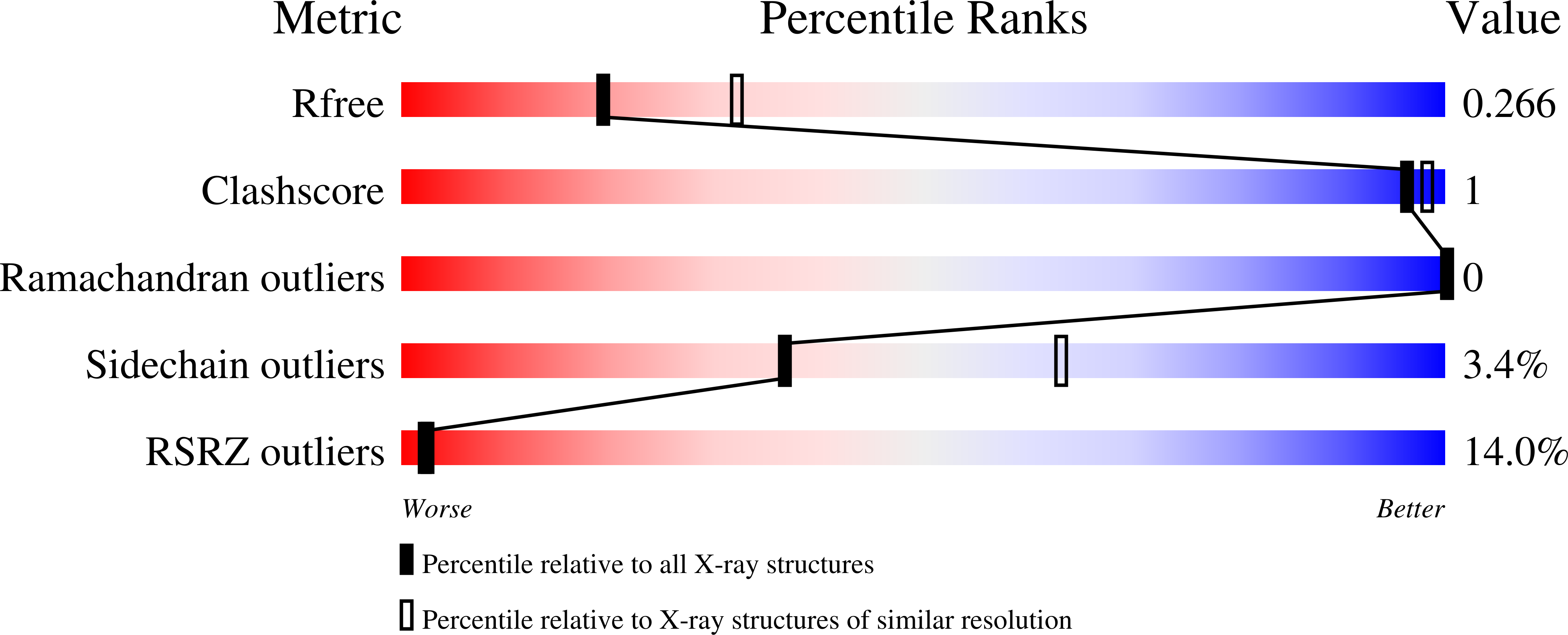
R-Value Free:
0.24
R-Value Work:
0.20
R-Value Observed:
0.20
Space Group:
C 1 2 1