

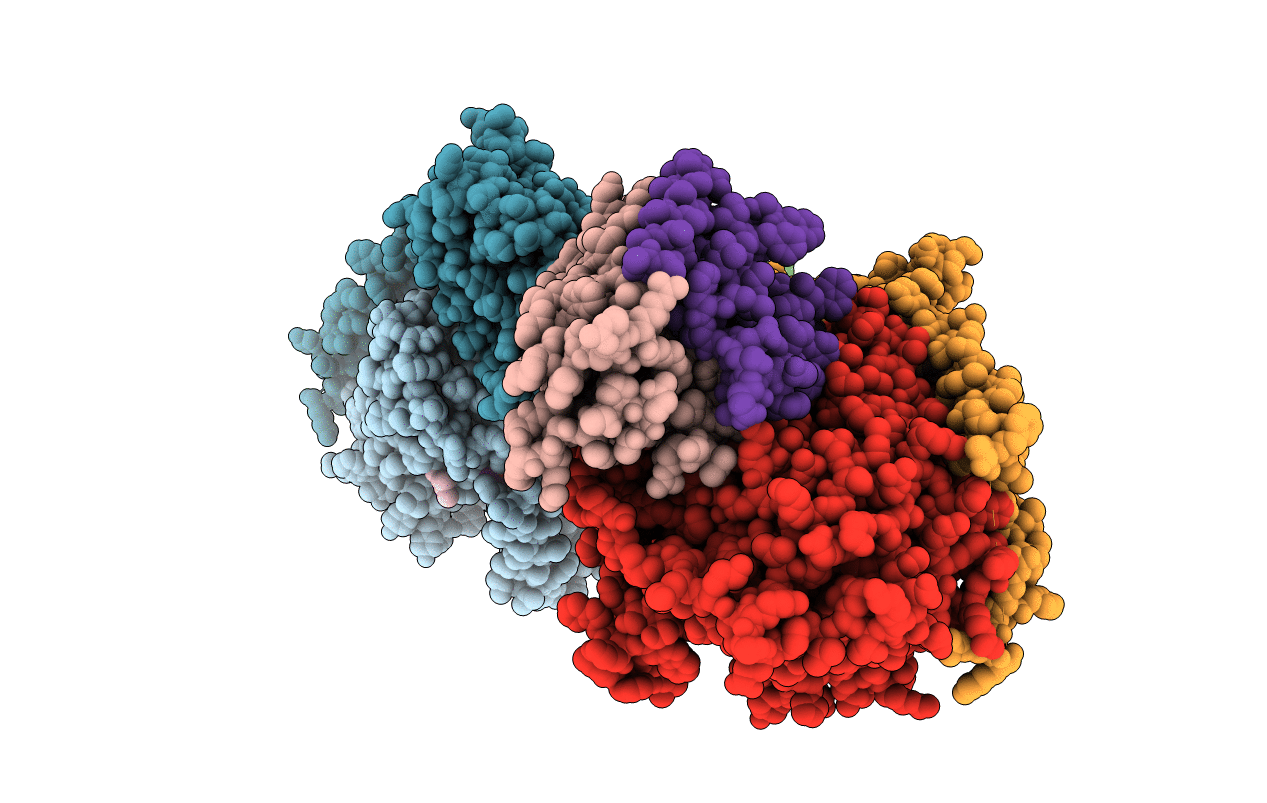
6KOC image
Deposition Date 2019-08-09
Release Date 2020-01-15
Last Version Date 2023-11-22
Entry Detail
PDB ID:
6KOC
Keywords:
Title:
X-ray Structure of the proton-pumping cytochrome aa3-600 menaquinol oxidase from Bacillus subtilis complexed with 3-iodo-N-oxo-2-heptyl-4-hydroxyquinoline
Biological Source:
Source Organism(s):
Expression System(s):
Method Details:
Experimental Method:
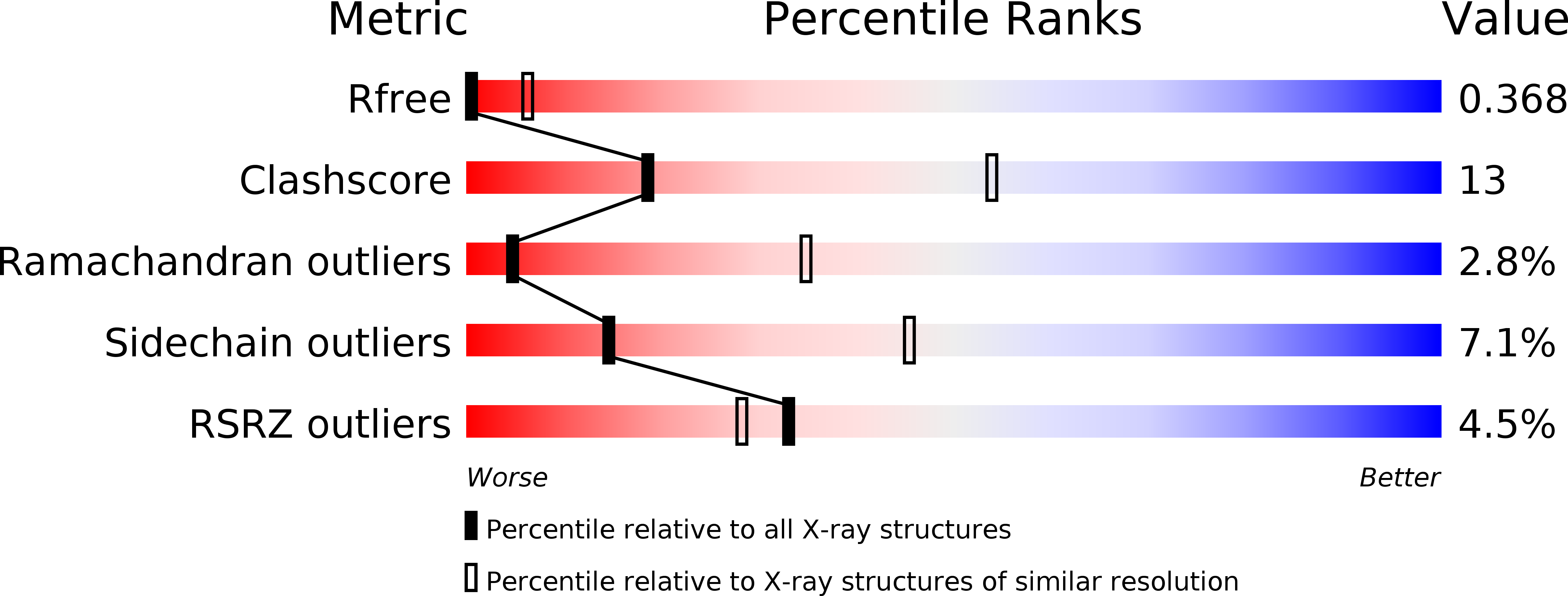
Resolution:
3.80 Å
R-Value Free:
0.36
R-Value Work:
0.32
Space Group:
P 1 21 1
Macromolecular Entities
Structures with similar UniProt ID
Protein Blast
Polymer Type:polypeptide(L)
Molecule:AA3-600 quinol oxidase subuni
Chain IDs:A, H (auth: E)
Chain Length:649
Number of Molecules:2
Biological Source:Bacillus subtilis
Structures with similar UniProt ID
Protein Blast
Polymer Type:polypeptide(L)
Molecule:Quinol oxidase subunit 2
Chain IDs:B, G (auth: F)
Chain Length:296
Number of Molecules:2
Biological Source:Bacillus subtilis
Structures with similar UniProt ID
Protein Blast
Polymer Type:polypeptide(L)
Molecule:AA3-600 quinol oxidase subuni
Chain IDs:C, E (auth: G)
Chain Length:204
Number of Molecules:2
Biological Source:Bacillus subtilis
Structures with similar UniProt ID
Protein Blast
Polymer Type:polypeptide(L)
Molecule:Quinol oxidase subunit 4,Quin
Gene (Uniprot):qoxD
Chain IDs:D, F (auth: H)
Chain Length:123
Number of Molecules:2
Biological Source:Bacillus subtilis subsp. subtilis str. 168, Bacillus subtilis (strain 168)
Primary Citation
Structure of the cytochrome
Proceedings of the National Academy of Sciences of the United States of America 117 872-876 ? (2020)
PMID: 31888984 DOI: .

Abstact

Virtually all proton-pumping terminal respiratory oxygen reductases are members of the heme-copper oxidoreductase superfamily. Most of these enzymes use reduced cytochrome c as a source of electrons, but a group of enzymes have evolved to directly oxidize membrane-bound quinols, usually menaquinol or ubiquinol. All of the quinol oxidases have an additional transmembrane helix (TM0) in subunit I that is not present in the related cytochrome c oxidases. The current work reports the 3.6-Å-resolution X-ray structure of the cytochrome aa3 -600 menaquinol oxidase from Bacillus subtilis containing 1 equivalent of menaquinone. The structure shows that TM0 forms part of a cleft to accommodate the menaquinol-7 substrate. Crystals which have been soaked with the quinol-analog inhibitor HQNO (N-oxo-2-heptyl-4-hydroxyquinoline) or 3-iodo-HQNO reveal a single binding site where the inhibitor forms hydrogen bonds to amino acid residues shown previously by spectroscopic methods to interact with the semiquinone state of menaquinone, a catalytic intermediate.

Legend

Protein

Chemical

Disease

Primary Citation of related structures
Feedback Form
Name
Email
Institute
Feedback