

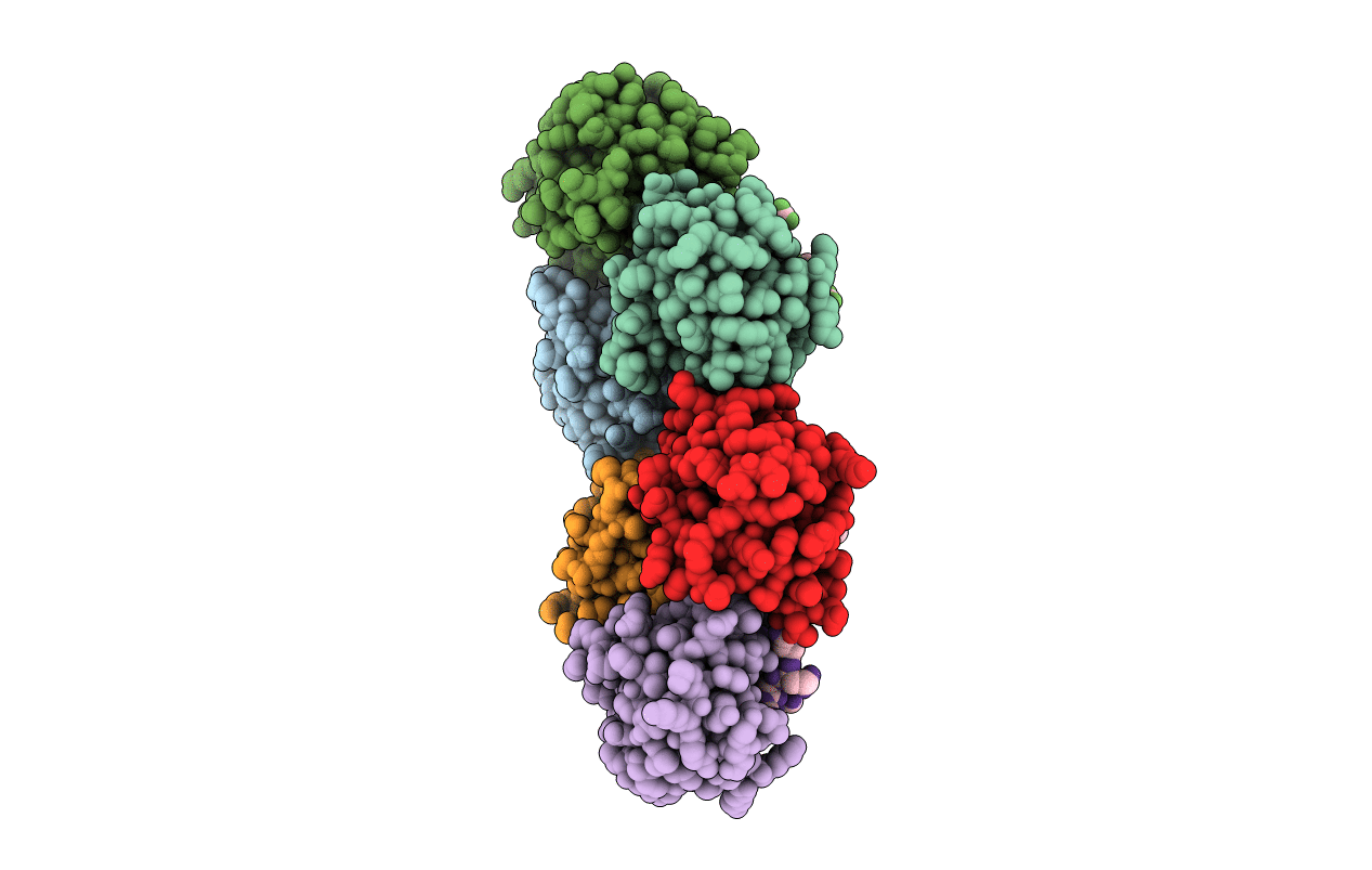
6KC4 image
Deposition Date 2019-06-27
Release Date 2019-09-04
Last Version Date 2024-10-16
Entry Detail
PDB ID:
6KC4
Title:
Crystal structure of human Fer SH2 domain bound to a phosphopeptide (DEpYENVD)
Biological Source:
Source Organism(s):
Expression System(s):
Method Details:
Experimental Method:
Resolution:
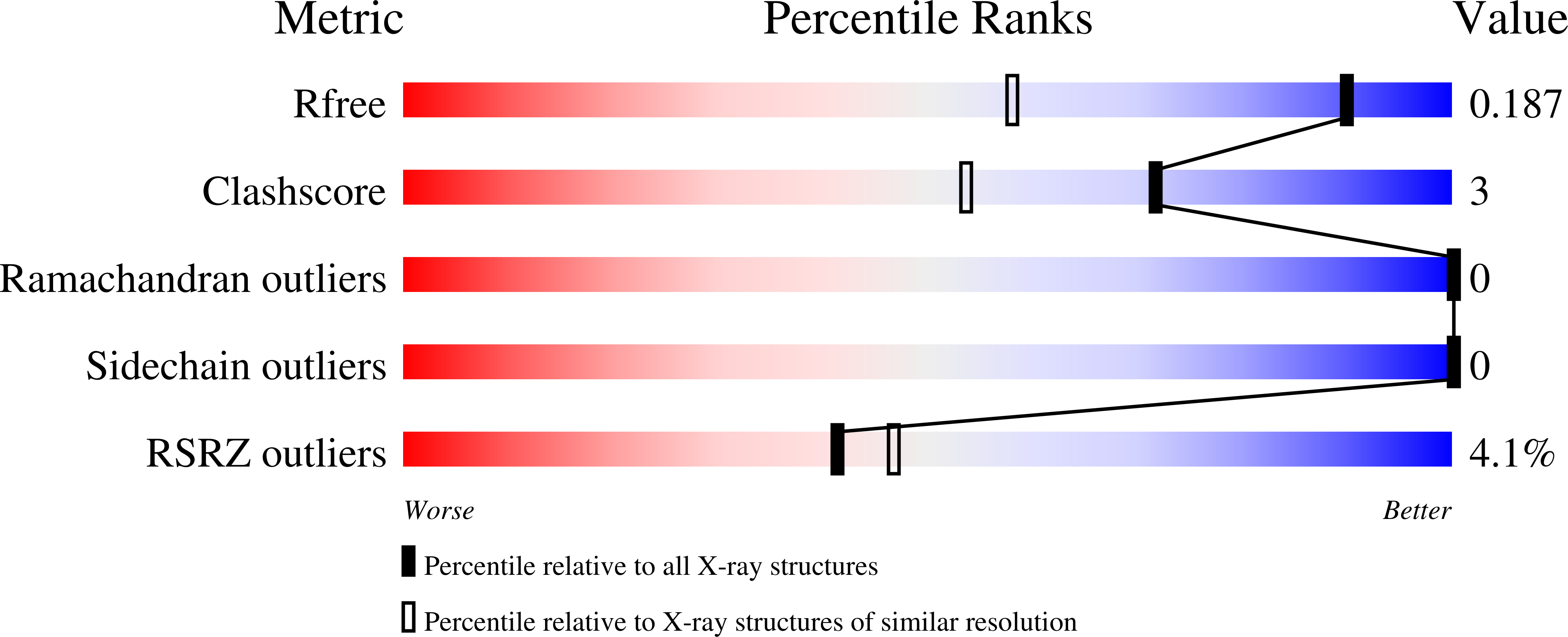
1.37 Å
R-Value Free:
0.18
R-Value Work:
0.16
R-Value Observed:
0.16
Space Group:
P 1 21 1
Macromolecular Entities
Polymer Type:polypeptide(L)
Molecule:Tyrosine-protein kinase Fer
Gene (Uniprot):FER
Chain IDs:A, C, E, G, I, K
Chain Length:102
Number of Molecules:6
Biological Source:Homo sapiens
Polymer Type:polypeptide(L)
Molecule:phosphopeptide (EDpYENVD)
Chain IDs:B, D, F, H, J, L
Chain Length:7
Number of Molecules:6
Biological Source:synthetic construct
Primary Citation
High-resolution structural analysis shows how different crystallographic environments can induce alternative modes of binding of a phosphotyrosine peptide to the SH2 domain of Fer tyrosine kinase.
Protein Sci. 28 2011 2019 (2019)
PMID: 31441171 DOI: 10.1002/pro.3713

Abstact

Fes and Fes-related (Fer) protein tyrosine kinases (PTKs) comprise a subfamily of nonreceptor tyrosine kinases characterized by a unique multidomain structure composed of an N-terminal Fer/CIP4 homology-Bin/Amphiphysin/Rvs (F-BAR) domain, a central Src homology 2 (SH2) domain, and a C-terminal PTK domain. Fer is ubiquitously expressed, and upregulation of Fer has been implicated in various human cancers. The PTK activity of Fes has been shown to be positively regulated by the binding of phosphotyrosine-containing ligands to the SH2 domain. Here, the X-ray crystal structure of human Fer SH2 domain bound to a phosphopeptide that has D-E-pY-E-N-V-D sequence is reported at 1.37 å resolution. The asymmetric unit (ASU) contains six Fer-phosphopeptide complexes, and the structure reveals three distinct binding modes for the same phosphopeptide. At four out of the six binding sites in the ASU, the phosphopeptide binds to Fer SH2 domain in a type I β-turn conformation, and this could be the optimal binding mode of this phosphopeptide. At the other two binding sites in the ASU, it appears that spatial proximity of neighboring SH2 domains in the crystal induces alternative modes of binding of this phosphopeptide.

Legend

Protein

Chemical

Disease

Primary Citation of related structures
Feedback Form
Name
Email
Institute
Feedback