Deposition Date
2018-06-21
Release Date
2018-08-22
Last Version Date
2025-09-17
Entry Detail
PDB ID:
6A4U
Keywords:
Title:
The first crystal structure of crustacean ferritin that is a hybrid type of H and L ferritin
Biological Source:
Source Organism(s):
Marsupenaeus japonicus (Taxon ID: 27405)
Expression System(s):
Method Details:
Experimental Method:
Resolution:
1.16 Å
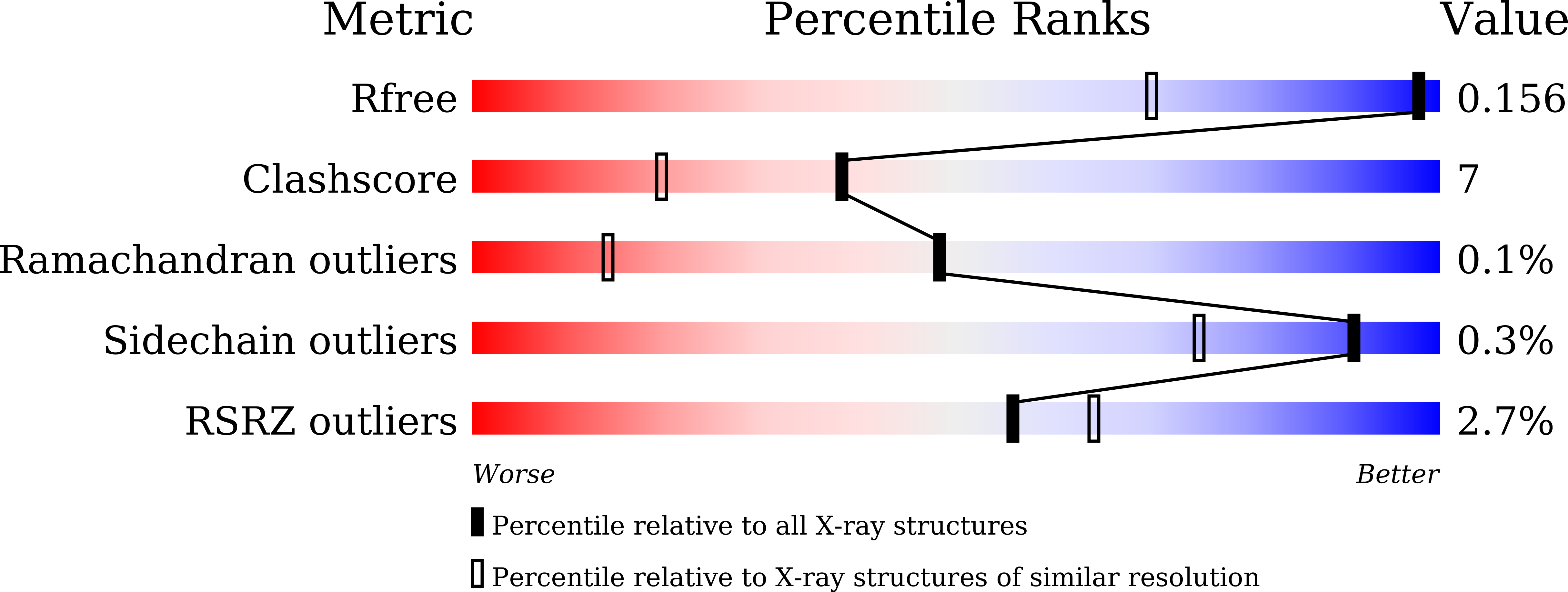
R-Value Free:
0.15
R-Value Work:
0.13
R-Value Observed:
0.13
Space Group:
I 4