5ZYC image
Deposition Date 2018-05-24
Release Date 2018-11-28
Last Version Date 2023-11-22
Entry Detail
PDB ID:
5ZYC
Keywords:
Title:
Crystal Structure of Glucose Isomerase Soaked with Mn2+
Biological Source:
Source Organism(s):
Expression System(s):
Method Details:
Experimental Method:
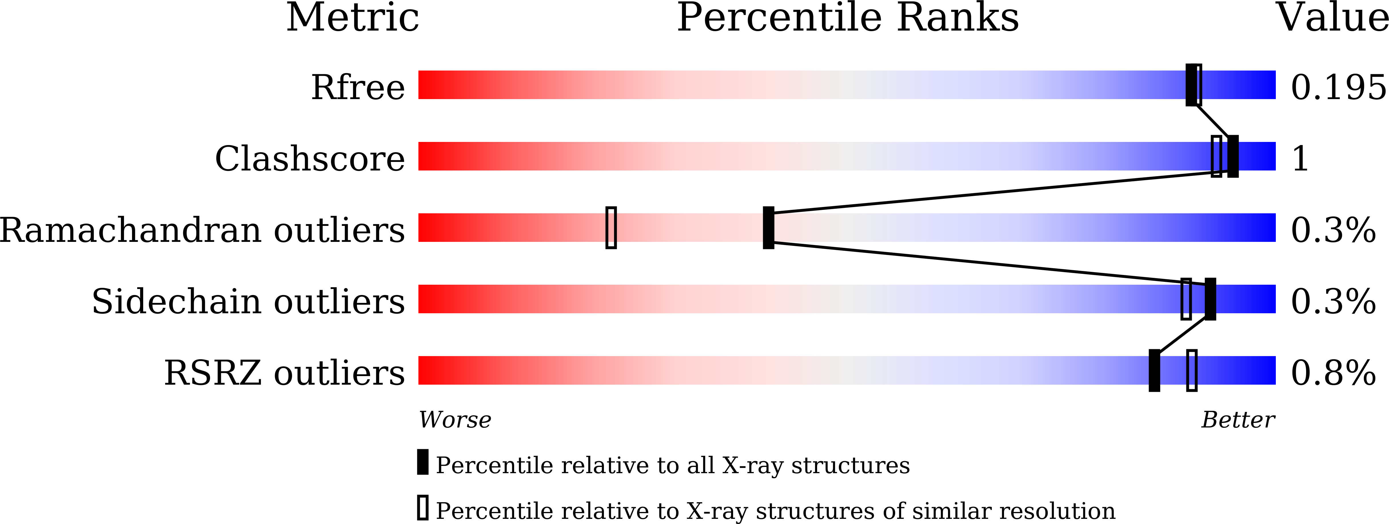
Resolution:
1.75 Å
R-Value Free:
0.18
R-Value Work:
0.16
R-Value Observed:
0.16
Space Group:
I 2 2 2
Macromolecular Entities
Structures with similar UniProt ID
Protein Blast
Polymer Type:polypeptide(L)
Molecule:Xylose isomerase
Gene (Uniprot):xylA
Chain IDs:A
Chain Length:388
Number of Molecules:1
Biological Source:Streptomyces rubiginosus
Primary Citation
Structural analysis of substrate recognition by glucose isomerase in Mn2+binding mode at M2 site in S. rubiginosus
Biochem. Biophys. Res. Commun. 503 770 775 (2018)
PMID: 29909012 DOI: 10.1016/j.bbrc.2018.06.074

Abstact

Glucose isomerase (GI) catalyzes the reversible enzymatic isomerization of d-glucose and d-xylose to d-fructose and d-xylulose, respectively. This is one of the most important enzymes in the production of high-fructose corn syrup (HFCS) and biofuel. We recently determined the crystal structure of GI from S. rubiginosus (SruGI) complexed with a xylitol inhibitor in one metal binding mode. Although we assessed inhibitor binding at the M1 site, the metal binding at the M2 site and the substrate recognition mechanism for SruGI remains the unclear. Here, we report the crystal structure of the two metal binding modes of SruGI and its complex with glucose. This study provides a snapshot of metal binding at the SruGI M2 site in the presence of Mn2+, but not in the presence of Mg2+. Metal binding at the M2 site elicits a configuration change at the M1 site. Glucose molecule can only bind to the M1 site in presence of Mn2+ at the M2 site. Glucose and Mn2+ at the M2 site were bridged by water molecules using a hydrogen bonding network. The metal binding geometry of the M2 site indicates a distorted octahedral coordination with an angle of 55-110°, whereas the M1 site has a relatively stable octahedral coordination with an angle of 85-95°. We suggest a two-step sequential process for SruGI substrate recognition, in Mn2+ binding mode, at the M2 site. Our results provide a better understanding of the molecular role of the M2 site in GI substrate recognition.

Legend

Protein

Chemical

Disease

Primary Citation of related structures
Feedback Form
Name
Email
Institute
Feedback