5YGM image
Deposition Date 2017-09-24
Release Date 2017-10-25
Last Version Date 2023-11-22
Entry Detail
PDB ID:
5YGM
Title:
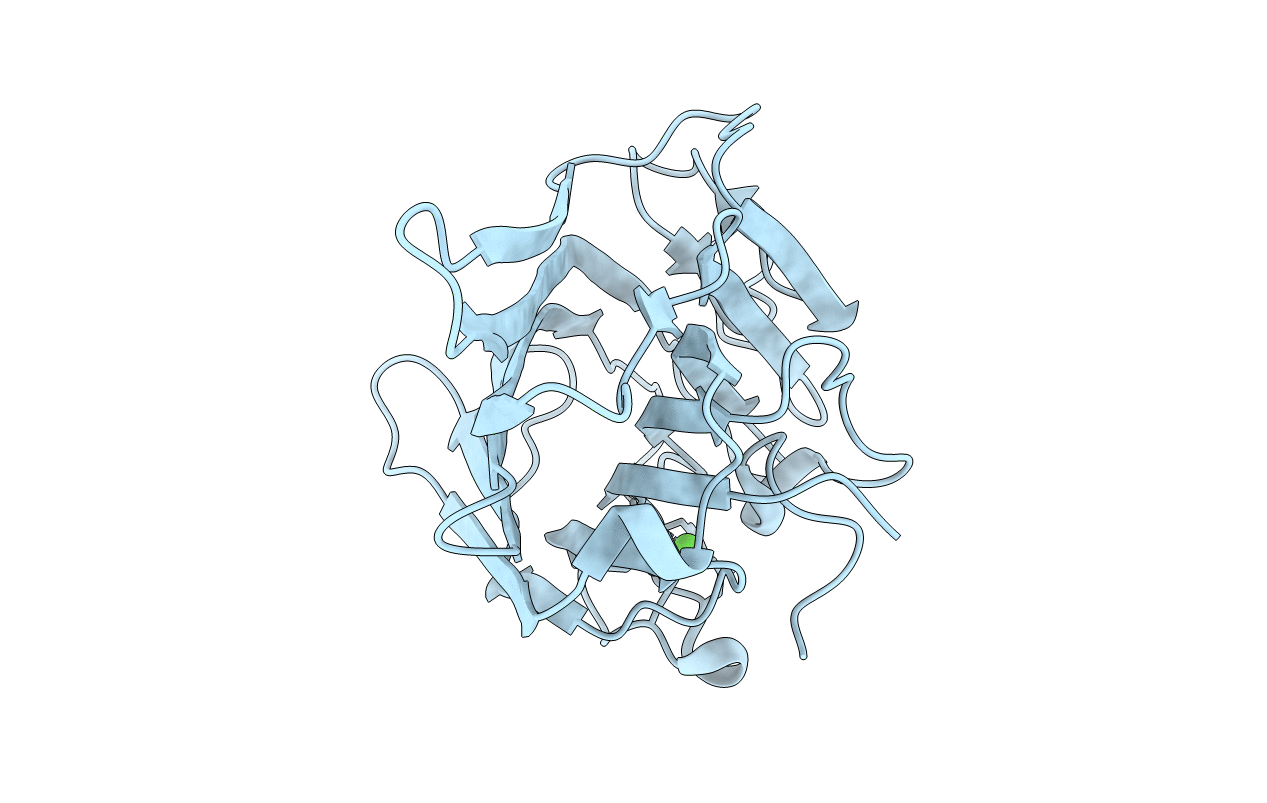
Monomeric structure of concanavalin A at pH 7.5 from Carnivalia ensiformis
Biological Source:
Source Organism(s):
Expression System(s):
Method Details:
Experimental Method:
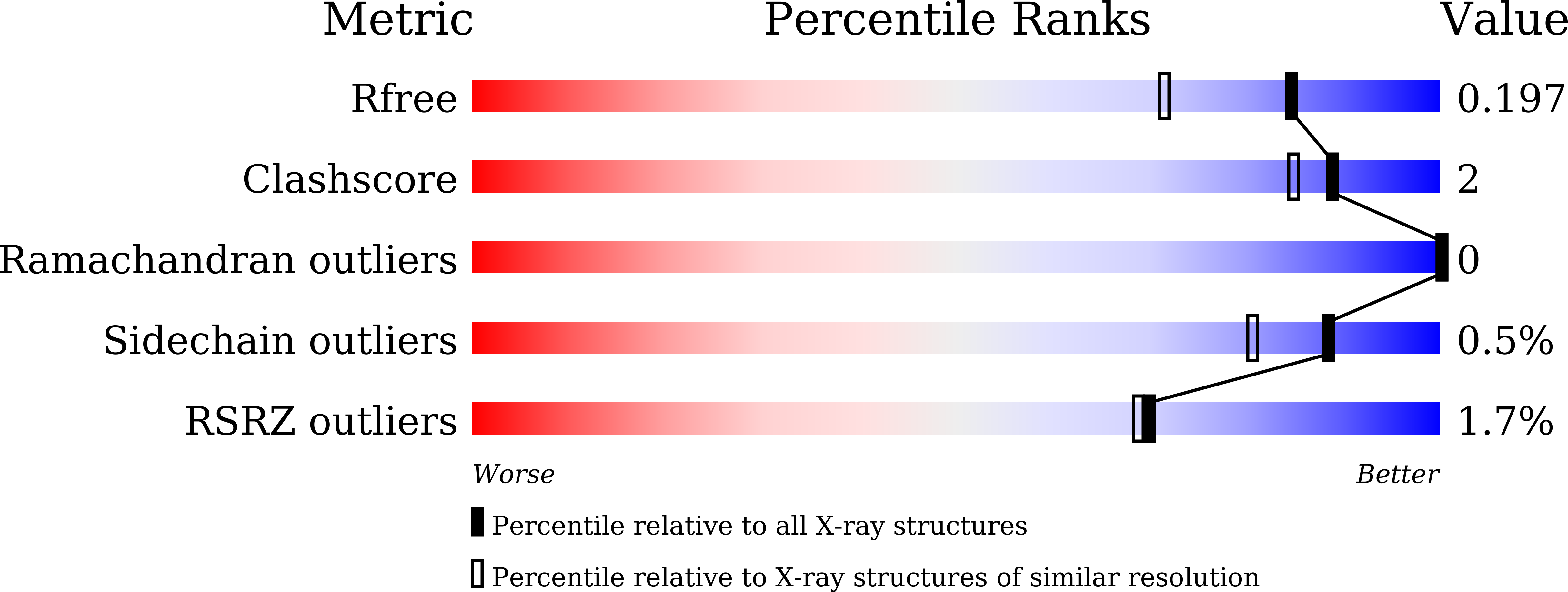
Resolution:
1.60 Å
R-Value Free:
0.19
R-Value Work:
0.16
R-Value Observed:
0.16
Space Group:
I 2 2 2
Macromolecular Entities
Structures with similar UniProt ID
Protein Blast
Polymer Type:polypeptide(L)
Molecule:Concanavalin-A
Chain IDs:A
Chain Length:237
Number of Molecules:1
Biological Source:Canavalia ensiformis
Primary Citation
Heterometal-Coordinated Monomeric Concanavalin A at pH 7.5 from Canavalia ensiformis
J. Microbiol. Biotechnol. 27 2241 2244 (2017)
PMID: 29025256 DOI: 10.4014/jmb.1709.09057

Abstact

The structure of concanavalin A (ConA) has been studied intensively owing to its specific interactions with carbohydrates and its heterometal (Ca²⁺ and Mn²⁺) coordination. Most structures from X-ray crystallography have shown ConA as a dimer or tetramer, because the complex formation requires specific crystallization conditions. Here, we reported the monomeric structure of ConA with a resolution of 1.6 Å, which revealed that metal coordination could trigger sugar-binding ability. The calcium coordination residue, Asn14, changed the orientation of carbohydrate-binding residues and biophysical details, including structural information, providing valuable clues for the development and application of detection kits using ConA.

Legend

Protein

Chemical

Disease

Primary Citation of related structures
Feedback Form
Name
Email
Institute
Feedback