5ES4 image
Deposition Date 2015-11-16
Release Date 2016-03-02
Last Version Date 2024-11-06
Entry Detail
PDB ID:
5ES4
Keywords:
Title:
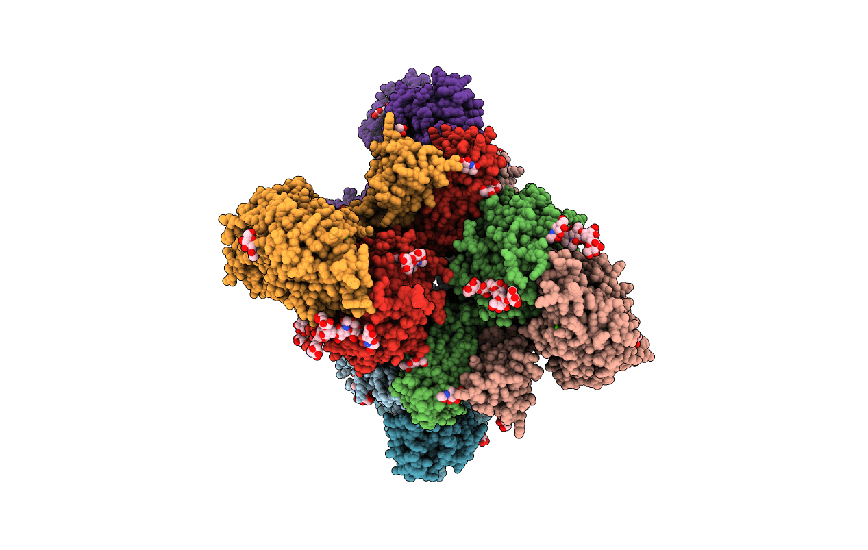
RE-REFINEMENT OF INTEGRIN ALPHAXBETA2 ECTODOMAIN IN THE CLOSED/BENT CONFORMATION
Biological Source:
Source Organism(s):
Homo sapiens (Taxon ID: 9606)
Expression System(s):
Method Details:
Experimental Method:
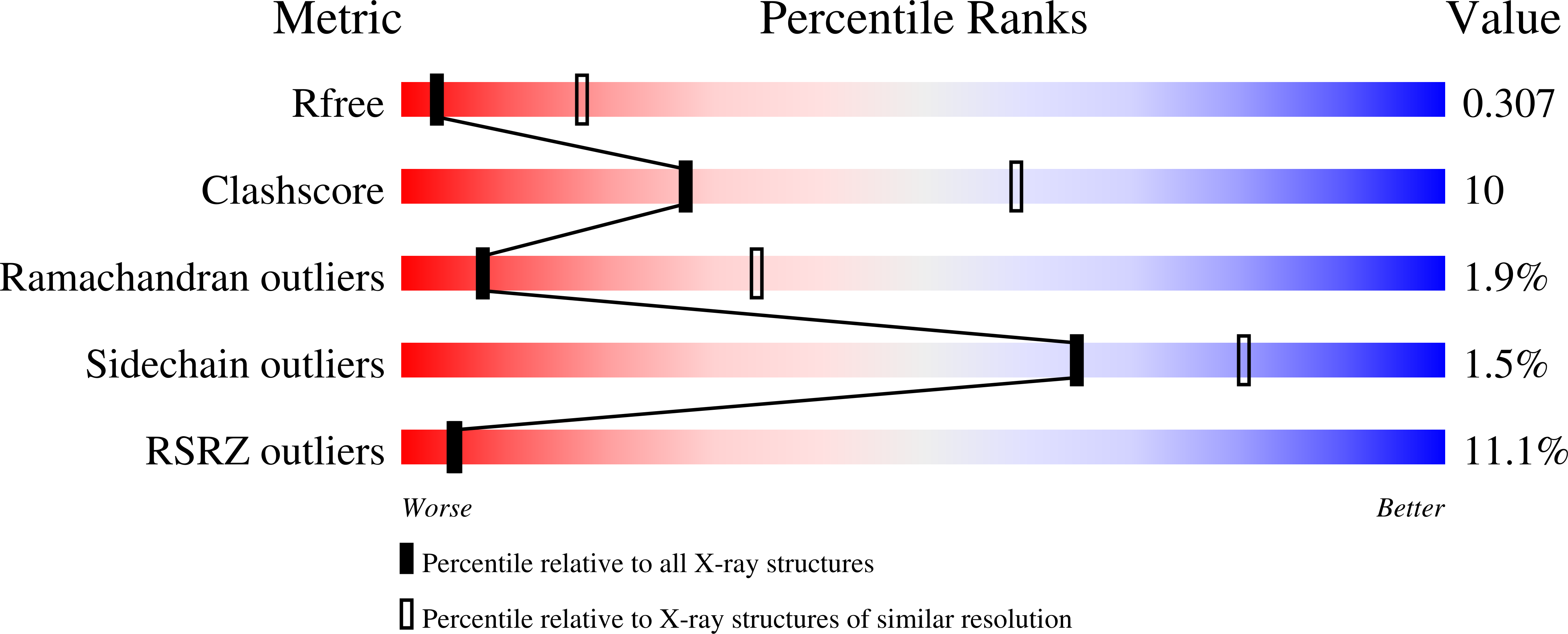
Resolution:
3.30 Å
R-Value Free:
0.30
R-Value Work:
0.25
R-Value Observed:
0.25
Space Group:
P 21 21 21
Macromolecular Entities
Structures with similar UniProt ID
Protein Blast
Polymer Type:polypeptide(L)
Molecule:Integrin alpha-X
Gene (Uniprot):ITGAX
Chain IDs:A, C, E, G
Chain Length:1137
Number of Molecules:4
Biological Source:Homo sapiens
Structures with similar UniProt ID
Protein Blast
Polymer Type:polypeptide(L)
Molecule:Integrin beta-2
Gene (Uniprot):ITGB2
Chain IDs:B, D, F, H
Chain Length:727
Number of Molecules:4
Biological Source:Homo sapiens
Primary Citation
Leukocyte integrin alpha L beta 2 headpiece structures: The alpha I domain, the pocket for the internal ligand, and concerted movements of its loops.
Proc. Natl. Acad. Sci. U.S.A. 113 2940 2945 (2016)
PMID: 26936951 DOI: 10.1073/pnas.1601379113

Abstact

High-resolution crystal structures of the headpiece of lymphocyte function-associated antigen-1 (integrin αLβ2) reveal how the αI domain interacts with its platform formed by the α-subunit β-propeller and β-subunit βI domains. The αLβ2 structures compared with αXβ2 structures show that the αI domain, tethered through its N-linker and a disulfide to a stable β-ribbon pillar near the center of the platform, can undergo remarkable pivoting and tilting motions that appear buffered by N-glycan decorations that differ between αL and αX subunits. Rerefined β2 integrin structures reveal details including pyroglutamic acid at the β2 N terminus and bending within the EGF1 domain. Allostery is relayed to the αI domain by an internal ligand that binds to a pocket at the interface between the β-propeller and βI domains. Marked differences between the αL and αX subunit β-propeller domains concentrate near the binding pocket and αI domain interfaces. Remarkably, movement in allostery in the βI domain of specificity determining loop 1 (SDL1) causes concerted movement of SDL2 and thereby tightens the binding pocket for the internal ligand.

Legend

Protein

Chemical

Disease

Primary Citation of related structures
Feedback Form
Name
Email
Institute
Feedback