Deposition Date
2015-06-12
Release Date
2016-01-06
Last Version Date
2024-11-06
Entry Detail
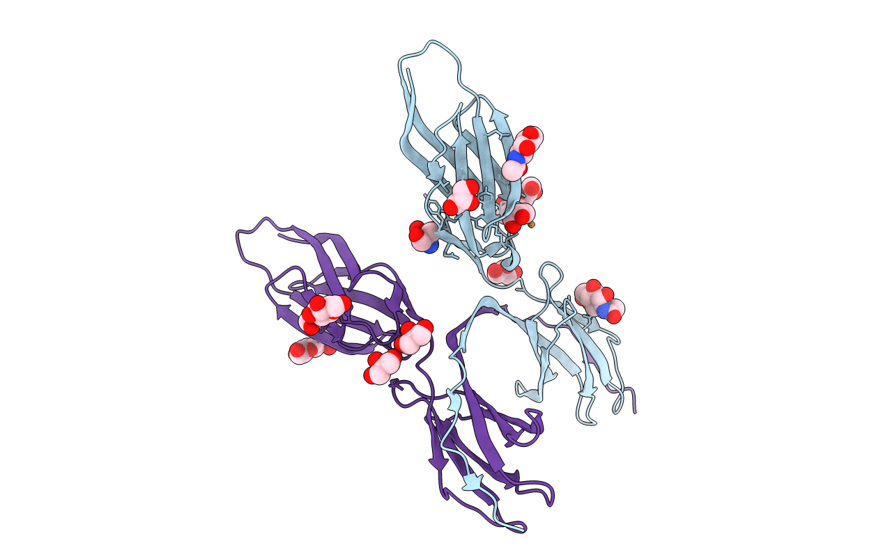
Biological Source:
Source Organism(s):
Homo sapiens (Taxon ID: 9606)
Expression System(s):
Method Details:
Experimental Method:
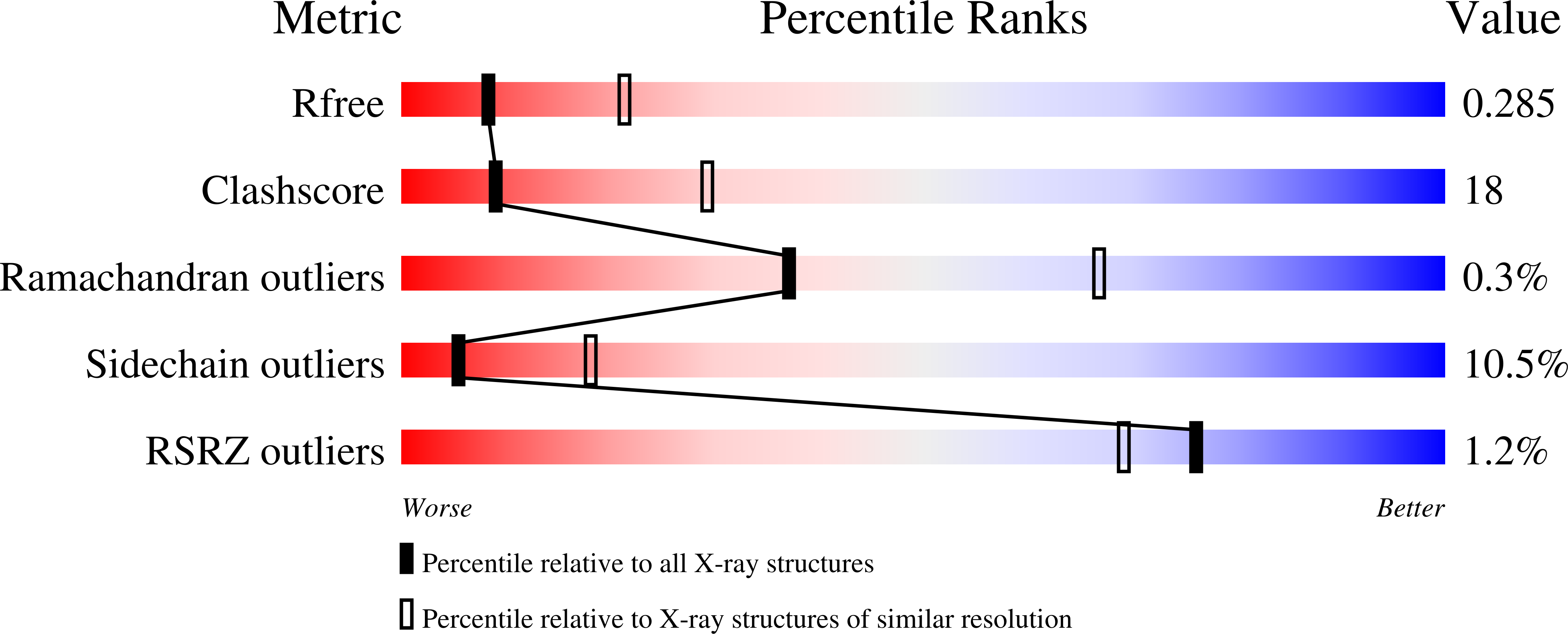
Resolution:
2.80 Å
R-Value Free:
0.27
R-Value Work:
0.23
R-Value Observed:
0.23
Space Group:
I 41 2 2