

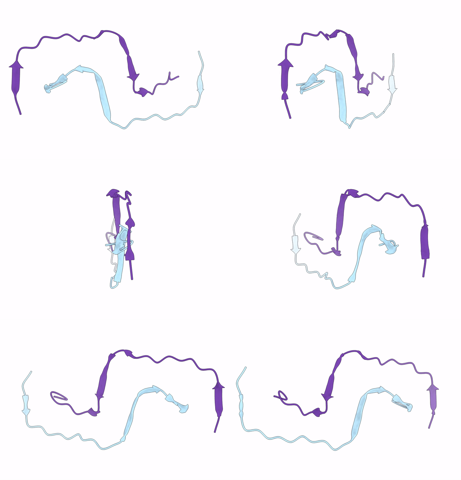
5AEF image
Deposition Date 2015-08-29
Release Date 2015-10-14
Last Version Date 2024-05-08
Entry Detail
PDB ID:
5AEF
Keywords:
Title:
Electron cryo-microscopy of an Abeta(1-42)amyloid fibril
Biological Source:
Source Organism(s):
HOMO SAPIENS (Taxon ID: 9606)
Method Details:
Experimental Method:
Resolution:
5.00 Å
Aggregation State:
FILAMENT
Reconstruction Method:
HELICAL
Macromolecular Entities
Polymer Type:polypeptide(L)
Molecule:AMYLOID BETA A4 PROTEIN
Chain IDs:A, B
Chain Length:28
Number of Molecules:2
Biological Source:HOMO SAPIENS
Ligand Molecules
Primary Citation
Peptide Dimer Structure in an Abeta(1-42) Fibril Visualized with Cryo-Em
Proc. Natl. Acad. Sci. U.S.A. 112 11858 ? (2015)
PMID: 26351699 DOI: 10.1073/PNAS.1503455112

Abstact

Alzheimer's disease (AD) is a fatal neurodegenerative disorder in humans and the main cause of dementia in aging societies. The disease is characterized by the aberrant formation of β-amyloid (Aβ) peptide oligomers and fibrils. These structures may damage the brain and give rise to cerebral amyloid angiopathy, neuronal dysfunction, and cellular toxicity. Although the connection between AD and Aβ fibrillation is extensively documented, much is still unknown about the formation of these Aβ aggregates and their structures at the molecular level. Here, we combined electron cryomicroscopy, 3D reconstruction, and integrative structural modeling methods to determine the molecular architecture of a fibril formed by Aβ(1-42), a particularly pathogenic variant of Aβ peptide. Our model reveals that the individual layers of the Aβ fibril are formed by peptide dimers with face-to-face packing. The two peptides forming the dimer possess identical tilde-shaped conformations and interact with each other by packing of their hydrophobic C-terminal β-strands. The peptide C termini are located close to the main fibril axis, where they produce a hydrophobic core and are surrounded by the structurally more flexible and charged segments of the peptide N termini. The observed molecular architecture is compatible with the general chemical properties of Aβ peptide and provides a structural basis for various biological observations that illuminate the molecular underpinnings of AD. Moreover, the structure provides direct evidence for a steric zipper within a fibril formed by full-length Aβ peptide.

Legend

Protein

Chemical

Disease

Primary Citation of related structures
Feedback Form
Name
Email
Institute
Feedback