Deposition Date
2015-04-21
Release Date
2015-06-10
Last Version Date
2024-10-23
Entry Detail
PDB ID:
4ZF3
Keywords:
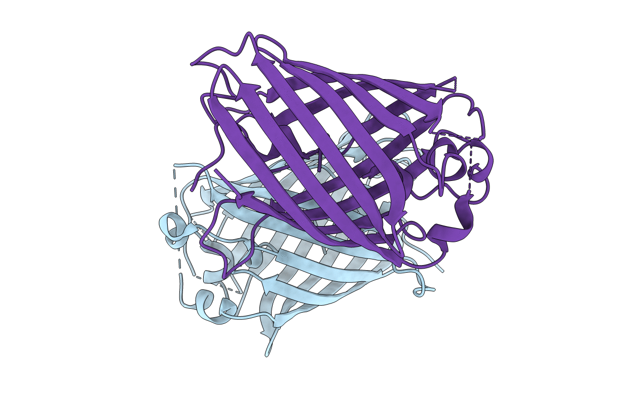
Title:
Crystal structure of Green Fluorescent Protein (GFP); S65T, H148D; circular permutant ( 50-51)
Biological Source:
Source Organism(s):
Aequorea victoria (Taxon ID: 6100)
Expression System(s):
Method Details:
Experimental Method:
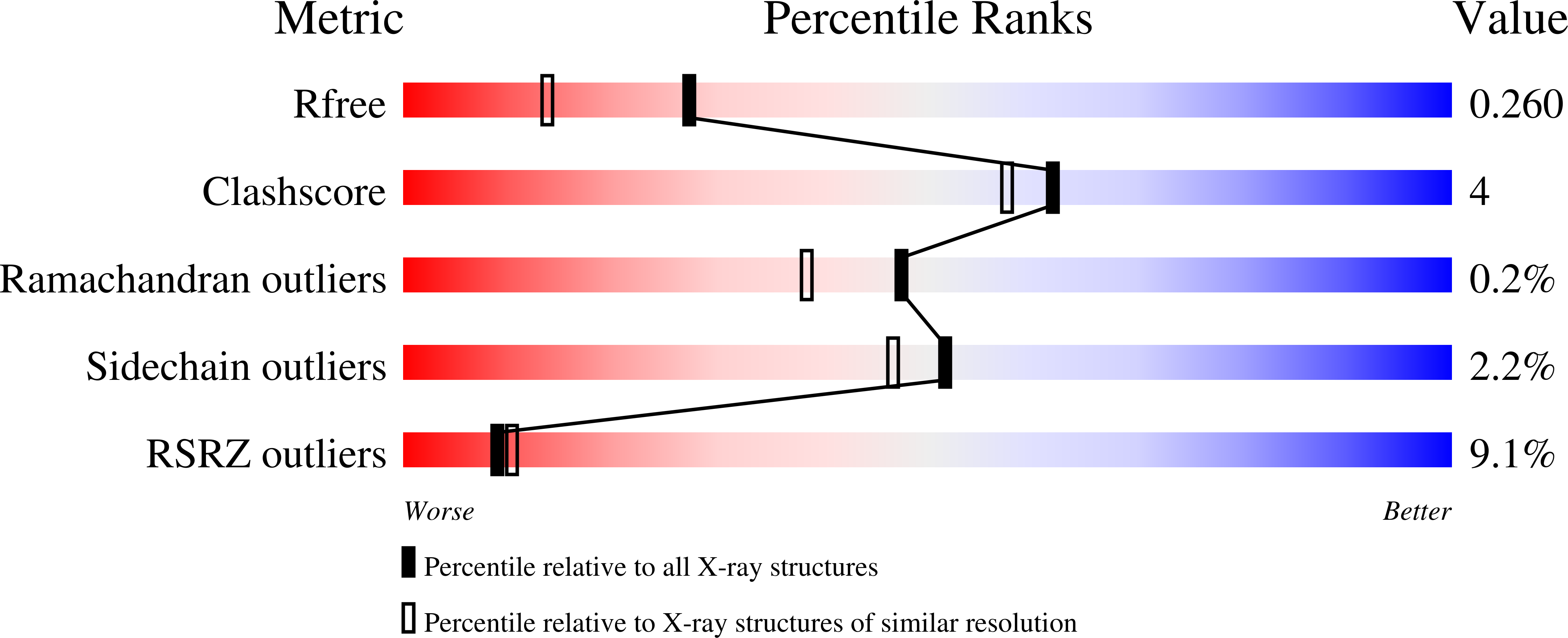
Resolution:
1.90 Å
R-Value Free:
0.26
R-Value Work:
0.21
R-Value Observed:
0.21
Space Group:
P 1 21 1