4Y97 image
Deposition Date 2015-02-17
Release Date 2015-04-15
Last Version Date 2023-09-27
Entry Detail
PDB ID:
4Y97
Keywords:
Title:
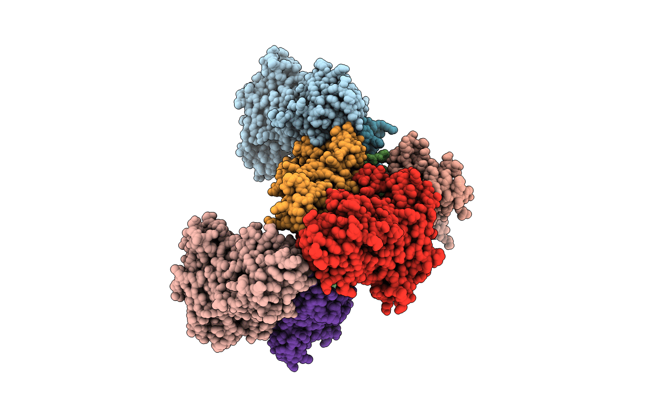
Crystal Structure of human Pol alpha B-subunit in complex with C-terminal domain of catalytic subunit
Biological Source:
Source Organism(s):
Homo sapiens (Taxon ID: 9606)
Expression System(s):
Method Details:
Experimental Method:
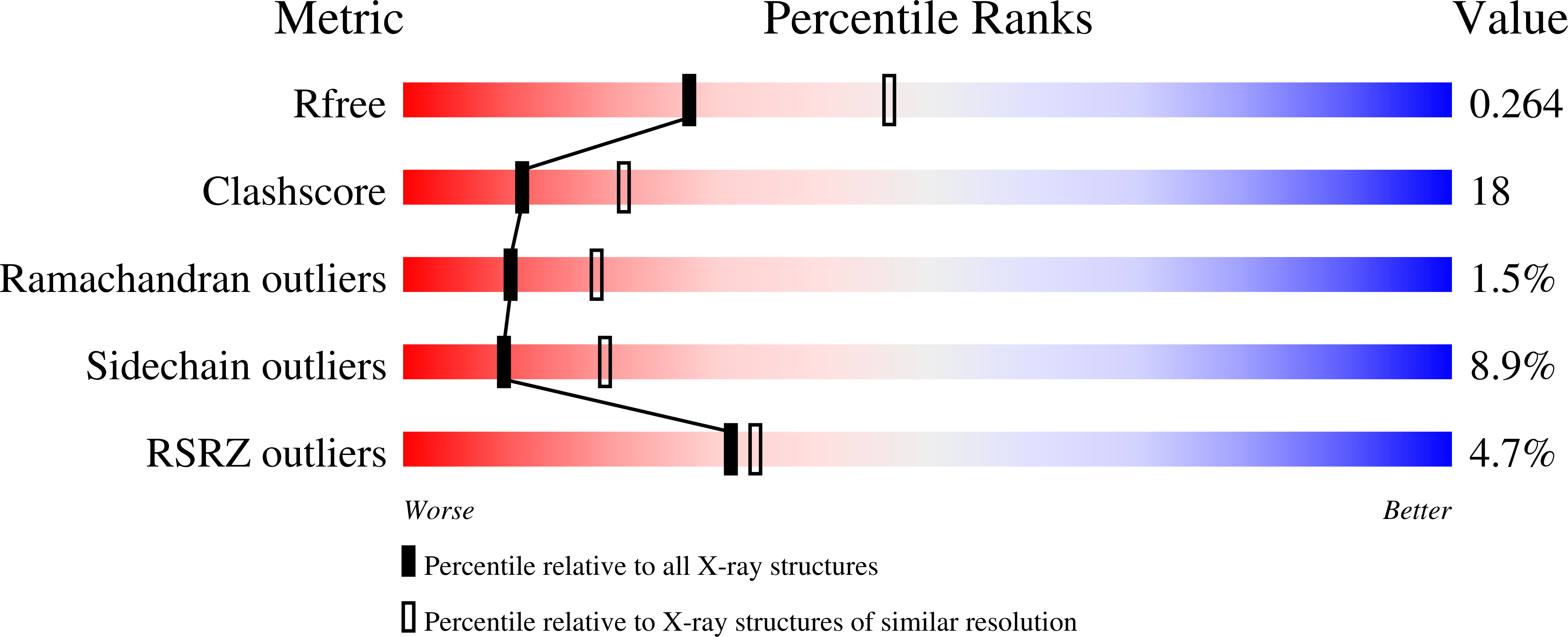
Resolution:
2.51 Å
R-Value Free:
0.25
R-Value Work:
0.21
R-Value Observed:
0.21
Space Group:
P 1 21 1
Macromolecular Entities
Structures with similar UniProt ID
Protein Blast
Polymer Type:polypeptide(L)
Molecule:DNA polymerase alpha subunit
Gene (Uniprot):POLA2
Chain IDs:A, C, E, G
Chain Length:598
Number of Molecules:4
Biological Source:Homo sapiens
Structures with similar UniProt ID
Protein Blast
Polymer Type:polypeptide(L)
Molecule:DNA polymerase alpha catalyti
Gene (Uniprot):POLA1
Chain IDs:B, D, F, H
Chain Length:180
Number of Molecules:4
Biological Source:Homo sapiens
Ligand Molecules
Primary Citation
Crystal Structure of the Human Pol alpha B Subunit in Complex with the C-terminal Domain of the Catalytic Subunit.
J. Biol. Chem. 290 14328 14337 (2015)
PMID: 25847248 DOI: 10.1074/jbc.M115.649954

Abstact

In eukaryotic DNA replication, short RNA-DNA hybrid primers synthesized by primase-DNA polymerase α (Prim-Pol α) are needed to start DNA replication by the replicative DNA polymerases, Pol δ and Pol ϵ. The C terminus of the Pol α catalytic subunit (p180C) in complex with the B subunit (p70) regulates the RNA priming and DNA polymerizing activities of Prim-Pol α. It tethers Pol α and primase, facilitating RNA primer handover from primase to Pol α. To understand these regulatory mechanisms and to reveal the details of human Pol α organization, we determined the crystal structure of p70 in complex with p180C. The structured portion of p70 includes a phosphodiesterase (PDE) domain and an oligonucleotide/oligosaccharide binding (OB) domain. The N-terminal domain and the linker connecting it to the PDE domain are disordered in the reported crystal structure. The p180C adopts an elongated asymmetric saddle shape, with a three-helix bundle in the middle and zinc-binding modules (Zn1 and Zn2) on each side. The extensive p180C-p70 interactions involve 20 hydrogen bonds and a number of hydrophobic interactions resulting in an extended buried surface of 4080 Å(2). Importantly, in the structure of the p180C-p70 complex with full-length p70, the residues from the N-terminal to the OB domain contribute to interactions with p180C. The comparative structural analysis revealed both the conserved features and the differences between the human and yeast Pol α complexes.

Legend

Protein

Chemical

Disease

Primary Citation of related structures
Feedback Form
Name
Email
Institute
Feedback