

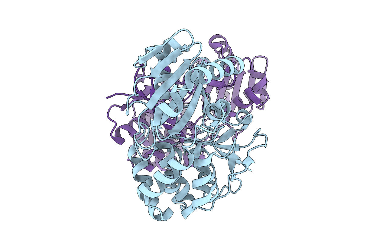
4RPA image
Deposition Date 2014-10-30
Release Date 2015-01-21
Last Version Date 2023-12-06
Entry Detail
PDB ID:
4RPA
Keywords:
Title:
Crystal structure of inorganic pyrophosphatase from Staphylococcus aureus in complex with Mn2+
Biological Source:
Source Organism(s):
Expression System(s):
Method Details:
Experimental Method:
Resolution:
2.10 Å
R-Value Free:
0.24
R-Value Work:
0.22
R-Value Observed:
0.22
Space Group:
P 61 2 2

Macromolecular Entities
Polymer Type:polypeptide(L)
Molecule:Probable manganese-dependent
Chain IDs:A, B
Chain Length:317
Number of Molecules:2
Biological Source:Staphylococcus aureus
Modified Residue
Compound ID Chain ID Parent Comp ID Details 2D Image
CSO A CYS S-HYDROXYCYSTEINE
OCS A CYS CYSTEINESULFONIC ACID
Ligand Molecules
Primary Citation
Structure of inorganic pyrophosphatase from Staphylococcus aureus reveals conformational flexibility of the active site.
J. Struct. Biol. 189 81 86 (2015)
PMID: 25576794 DOI: 10.1016/j.jsb.2014.12.003

Abstact

Cytoplasmic inorganic pyrophosphatase (PPiase) is an enzyme essential for survival of organisms, from bacteria to human. PPiases are divided into two structurally distinct families: family I PPiases are Mg(2+)-dependent and present in most archaea, eukaryotes and prokaryotes, whereas the relatively less understood family II PPiases are Mn(2+)-dependent and present only in some archaea, bacteria and primitive eukaryotes. Staphylococcus aureus (SA), a dangerous pathogen and a frequent cause of hospital infections, contains a family II PPiase (PpaC), which is an attractive potential target for development of novel antibacterial agents. We determined a crystal structure of SA PpaC in complex with catalytic Mn(2+) at 2.1Å resolution. The active site contains two catalytic Mn(2+) binding sites, each half-occupied, reconciling the previously observed 1:1 Mn(2+):enzyme stoichiometry with the presence of two divalent metal ion sites in the apo-enzyme. Unexpectedly, despite the absence of the substrate or products in the active site, the two domains of SA PpaC form a closed active site, a conformation observed in structures of other family II PPiases only in complex with substrate or product mimics. A region spanning residues 295-298, which contains a conserved substrate binding RKK motif, is flipped out of the active site, an unprecedented conformation for a PPiase. Because the mutant of Arg295 to an alanine is devoid of activity, this loop likely undergoes an induced-fit conformational change upon substrate binding and product dissociation. This closed conformation of SA PPiase may serve as an attractive target for rational design of inhibitors of this enzyme.

Legend

Protein

Chemical

Disease

Primary Citation of related structures
Feedback Form
Name
Email
Institute
Feedback