

4Q65 image
Deposition Date 2014-04-21
Release Date 2014-08-13
Last Version Date 2024-05-29
Entry Detail
PDB ID:
4Q65
Title:
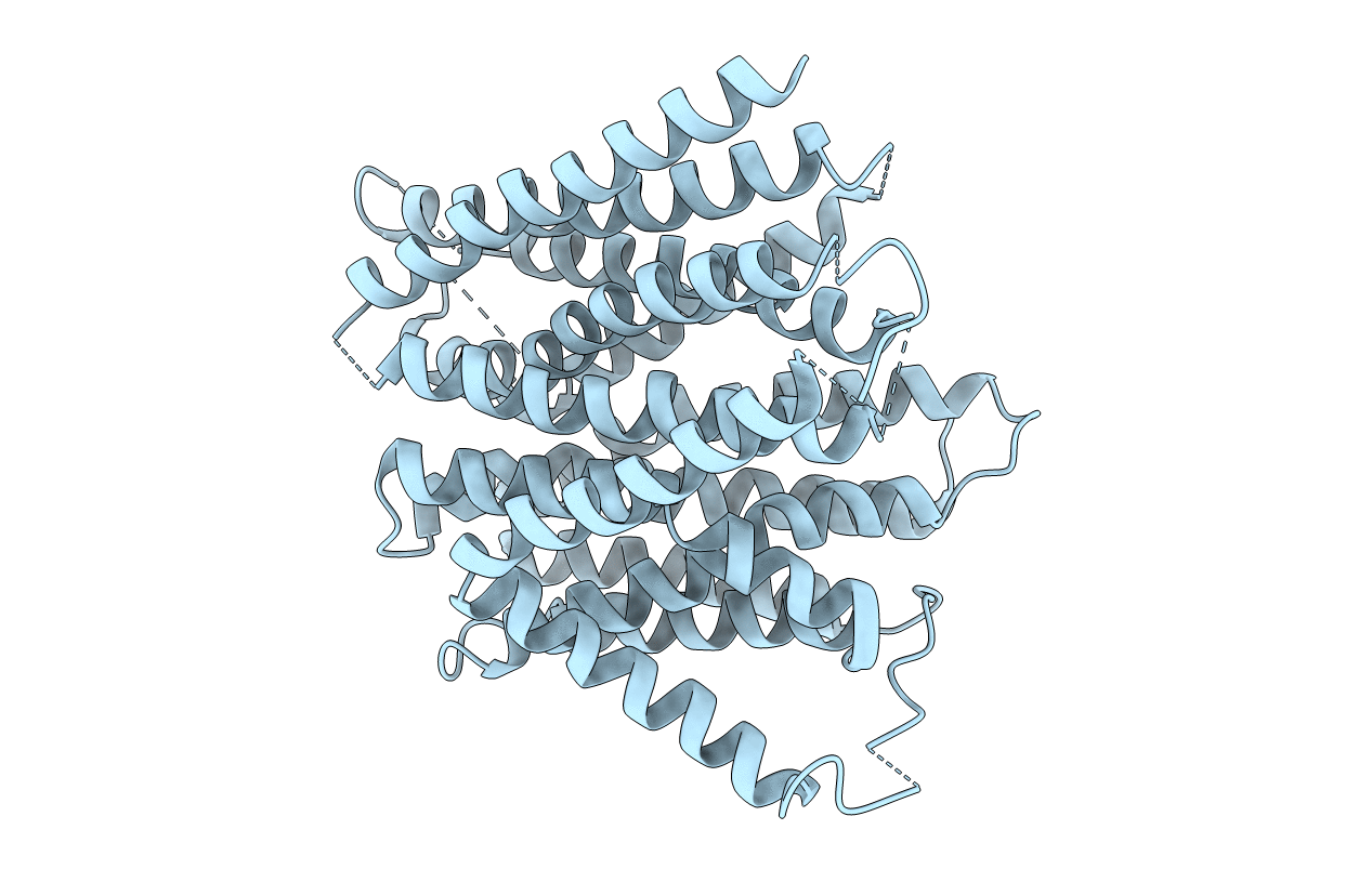
Structure of the E. coli Peptide Transporter YbgH
Biological Source:
Source Organism(s):
Expression System(s):
Method Details:
Experimental Method:
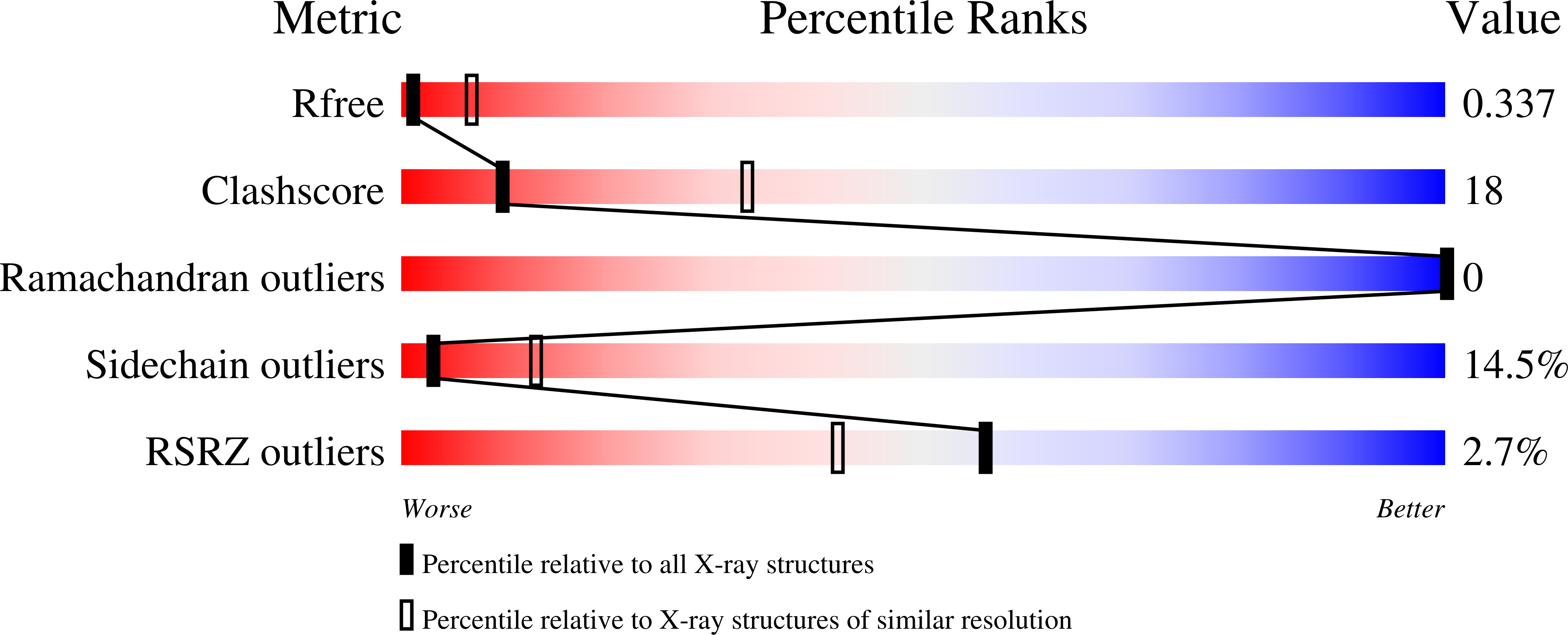
Resolution:
3.40 Å
R-Value Free:
0.31
R-Value Work:
0.25
R-Value Observed:
0.26
Space Group:
P 21 21 2
Macromolecular Entities
Polymer Type:polypeptide(L)
Molecule:Dipeptide permease D
Gene (Uniprot):dtpD
Mutagens:L472V
Chain IDs:A
Chain Length:493
Number of Molecules:1
Biological Source:Escherichia coli
Ligand Molecules
Primary Citation
Crystal structure of the E. coli peptide transporter YbgH.
Structure 22 1152 ? (2014)
PMID: 25066136 DOI: .

Abstact

E. coli YbgH belongs to the family of proton-dependent oligopeptide transporters (POTs), a subfamily of the major facilitator superfamily (MFS) of secondary active transporters. Like other MFS transporters, POT proteins switch between two major conformations during substrate transport. Apart from possessing a canonical 12-helix, two-domain transmembrane (TM) core, prokaryotic POT proteins usually have two TM helices inserted between the two domains. Here we determined the crystal structure of YbgH in its inward-facing conformation. Our structure-based functional studies investigated the roles of both the POT signature motif 2 and the inserted interdomain TM helix pair in the stabilization and regulation of the major conformational change in MFS/POT transporters. Furthermore, of all the proton-titratable amino acid residues, Glu21 is the only conserved one (among POTs) located in the central cavity and is critical for in vivo transport. Together, our results support the notion that MFS symporters utilize a transport mechanism based on substrate-protonation coupling.

Legend

Protein

Chemical

Disease

Primary Citation of related structures
Feedback Form
Name
Email
Institute
Feedback