4O63 image
Deposition Date 2013-12-20
Release Date 2014-09-24
Last Version Date 2023-09-20
Entry Detail
PDB ID:
4O63
Title:
Co-enzyme Induced Conformational Changes in Bovine Eye Glyceraldehyde 3-Phosphate Dehydrogenase
Biological Source:
Source Organism(s):
Bos taurus (Taxon ID: 9913)
Method Details:
Experimental Method:
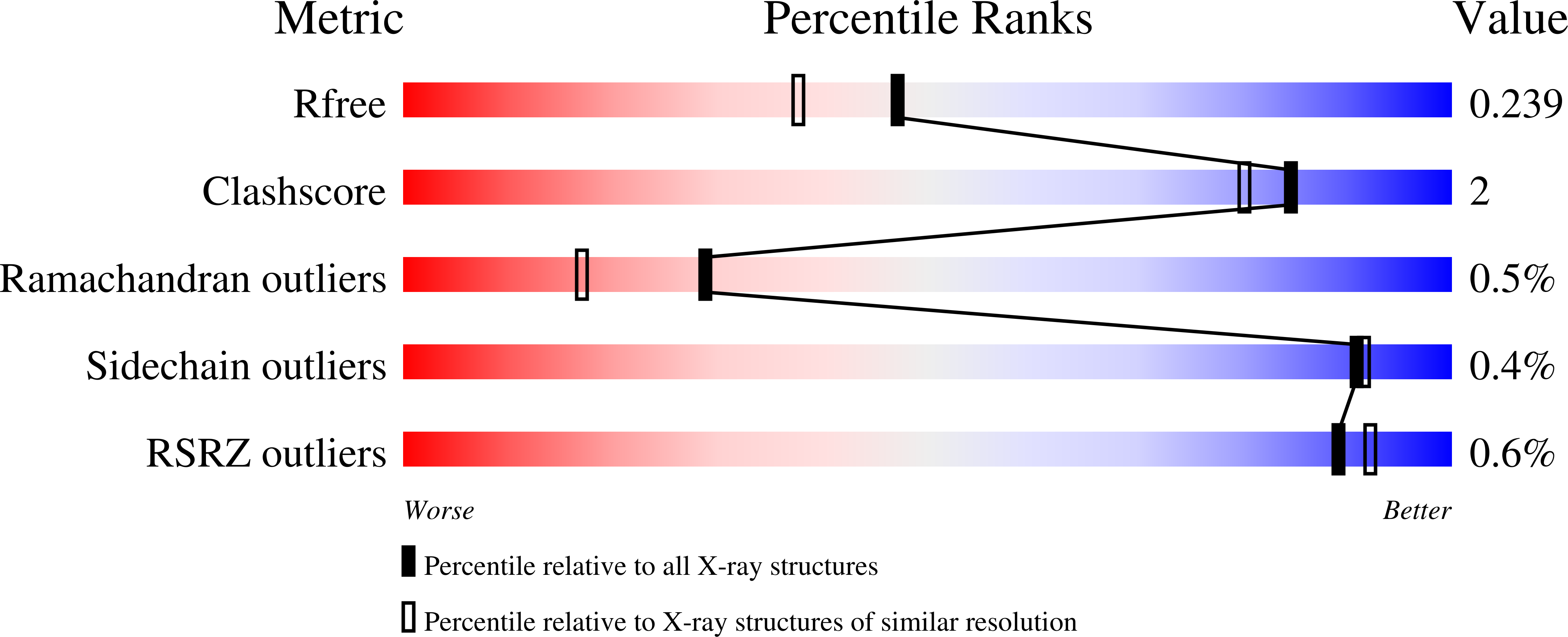
Resolution:
1.93 Å
R-Value Free:
0.23
R-Value Work:
0.18
R-Value Observed:
0.19
Space Group:
P 1 21 1
Macromolecular Entities
Structures with similar UniProt ID
Protein Blast
Polymer Type:polypeptide(L)
Molecule:Glyceraldehyde-3-phosphate de
Gene (Uniprot):GAPDH
Chain IDs:A (auth: P), B (auth: Q), C (auth: R), D (auth: O)
Chain Length:332
Number of Molecules:4
Biological Source:Bos taurus
Ligand Molecules
Primary Citation
High-resolution crystal structures of the photoreceptor glyceraldehyde 3-phosphate dehydrogenase (GAPDH) with three and four-bound NAD molecules.
Protein Sci. 23 1629 1639 (2014)
PMID: 25176140 DOI: 10.1002/pro.2543

Abstact

Glyceraldehyde-3-phosphate dehydrogenase (GAPDH) catalyzes the oxidative phosphorylation of d-glyceraldehyde 3-phosphate (G3P) into 1,3-diphosphoglycerate (BGP) in the presence of the NAD cofactor. GAPDH is an important drug target because of its central role in glycolysis, and nonglycolytic processes such as nuclear RNA transport, DNA replication/repair, membrane fusion and cellular apoptosis. Recent studies found that GAPDH participates in the development of diabetic retinopathy and its progression after the cessation of hyperglycemia. Here, we report two structures for native bovine photoreceptor GAPDH as a homotetramer with differing occupancy by NAD, bGAPDH(NAD)4 , and bGAPDH(NAD)3 . The bGAPDH(NAD)4 was solved at 1.52 Å, the highest resolution for GAPDH. Structural comparison of the bGAPDH(NAD)4 and bGAPDH(NAD)3 models revealed novel details of conformational changes induced by cofactor binding, including a loop region (residues 54-56). Structure analysis of bGAPDH confirmed the importance of Phe34 in NAD binding, and demonstrated that Phe34 was stabilized in the presence of NAD but displayed greater mobility in its absence. The oxidative state of the active site Cys149 residue is regulated by NAD binding, because this residue was found oxidized in the absence of dinucleotide. The distance between Cys149 and His176 decreased upon NAD binding and Cys149 remained in a reduced state when NAD was bound. These findings provide an important structural step for understanding the mechanism of GAPDH activity in vision and its pathological role in retinopathies.

Legend

Protein

Chemical

Disease

Primary Citation of related structures
Feedback Form
Name
Email
Institute
Feedback