

4MYA image
Deposition Date 2013-09-27
Release Date 2014-01-01
Last Version Date 2026-03-25
Entry Detail
PDB ID:
4MYA
Title:
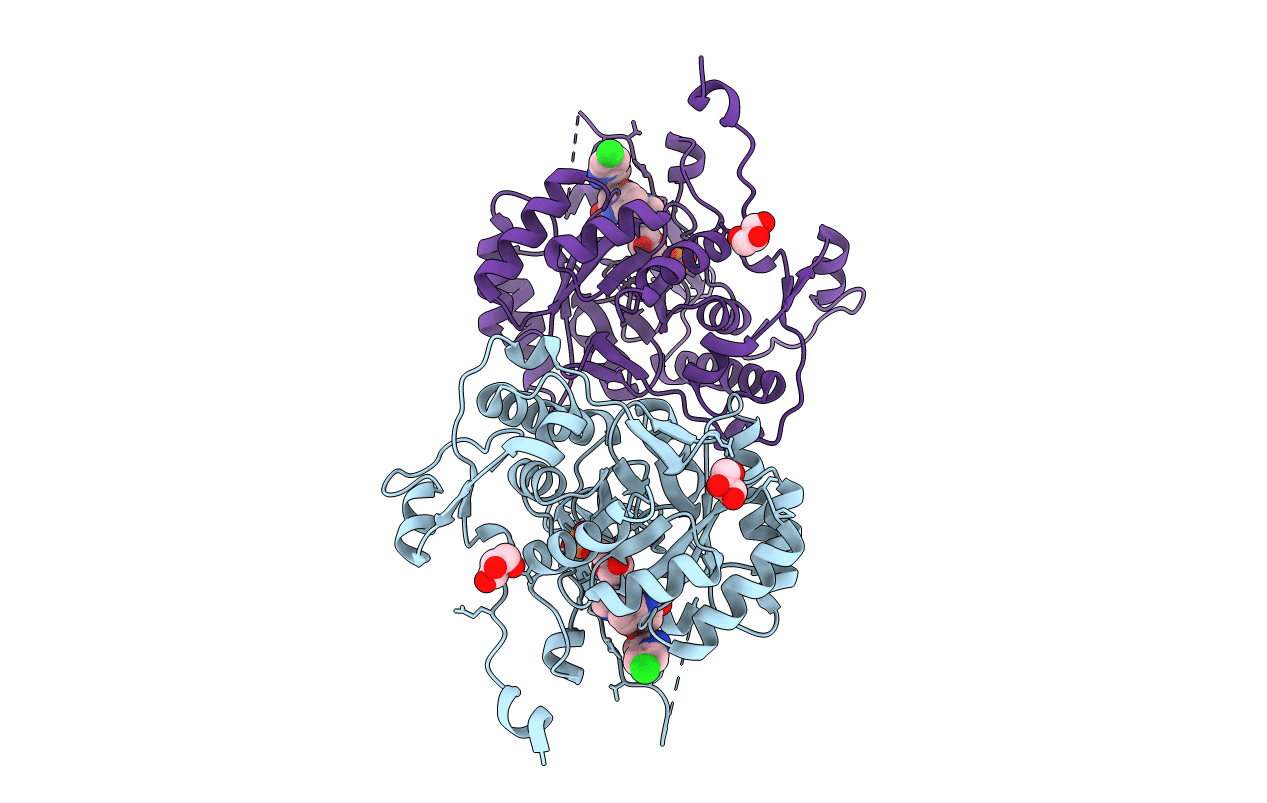
Crystal Structure of the Inosine 5'-monophosphate Dehydrogenase with an Internal Deletion of the CBS Domain from Bacillus anthracis str. Ames complexed with inhibitor A110
Biological Source:
Source Organism(s):
Expression System(s):
Method Details:
Experimental Method:
Resolution:
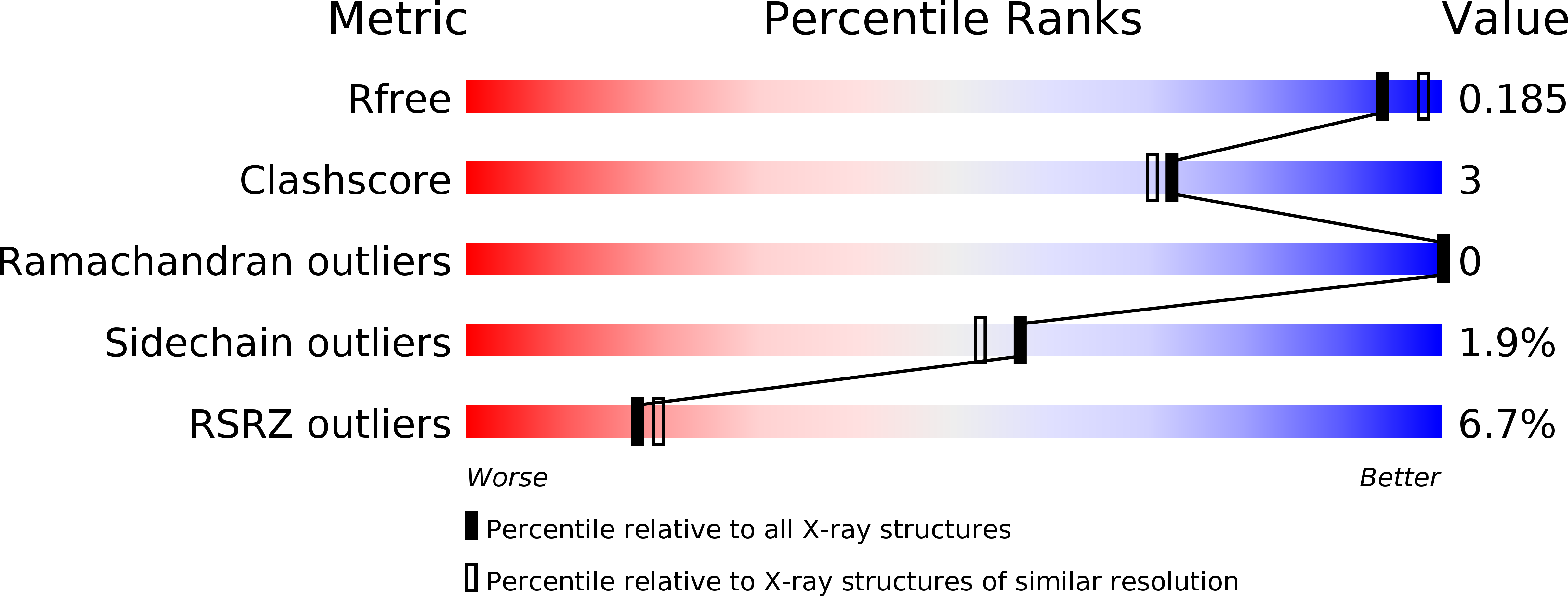
1.90 Å
R-Value Free:
0.18
R-Value Work:
0.14
R-Value Observed:
0.14
Space Group:
P 4
Macromolecular Entities
Structures with similar UniProt ID
Protein Blast
Polymer Type:polypeptide(L)
Molecule:Inosine-5'-monophosphate dehy
Mutagens:replaced residues 92-220 with GG
Chain IDs:A, B
Chain Length:384
Number of Molecules:2
Biological Source:Bacillus anthracis
Primary Citation
A novel cofactor-binding mode in bacterial IMP dehydrogenases explains inhibitor selectivity.
J. Biol. Chem. 290 5893 5911 (2015)
PMID: 25572472 DOI: 10.1074/jbc.M114.619767

Abstact

The steadily rising frequency of emerging diseases and antibiotic resistance creates an urgent need for new drugs and targets. Inosine 5'-monophosphate dehydrogenase (IMP dehydrogenase or IMPDH) is a promising target for the development of new antimicrobial agents. IMPDH catalyzes the oxidation of IMP to XMP with the concomitant reduction of NAD(+), which is the pivotal step in the biosynthesis of guanine nucleotides. Potent inhibitors of bacterial IMPDHs have been identified that bind in a structurally distinct pocket that is absent in eukaryotic IMPDHs. The physiological role of this pocket was not understood. Here, we report the structures of complexes with different classes of inhibitors of Bacillus anthracis, Campylobacter jejuni, and Clostridium perfringens IMPDHs. These structures in combination with inhibition studies provide important insights into the interactions that modulate selectivity and potency. We also present two structures of the Vibrio cholerae IMPDH in complex with IMP/NAD(+) and XMP/NAD(+). In both structures, the cofactor assumes a dramatically different conformation than reported previously for eukaryotic IMPDHs and other dehydrogenases, with the major change observed for the position of the NAD(+) adenosine moiety. More importantly, this new NAD(+)-binding site involves the same pocket that is utilized by the inhibitors. Thus, the bacterial IMPDH-specific NAD(+)-binding mode helps to rationalize the conformation adopted by several classes of prokaryotic IMPDH inhibitors. These findings offer a potential strategy for further ligand optimization.

Legend

Protein

Chemical

Disease

Primary Citation of related structures
Feedback Form
Name
Email
Institute
Feedback