

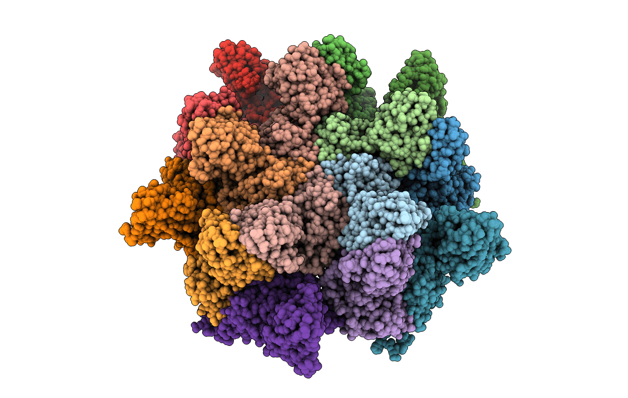
4LLF image
Deposition Date 2013-07-09
Release Date 2013-10-30
Last Version Date 2023-09-20
Entry Detail
PDB ID:
4LLF
Keywords:
Title:
Crystal structure of Cucumber Necrosis Virus
Biological Source:
Source Organism(s):
Method Details:
Experimental Method:
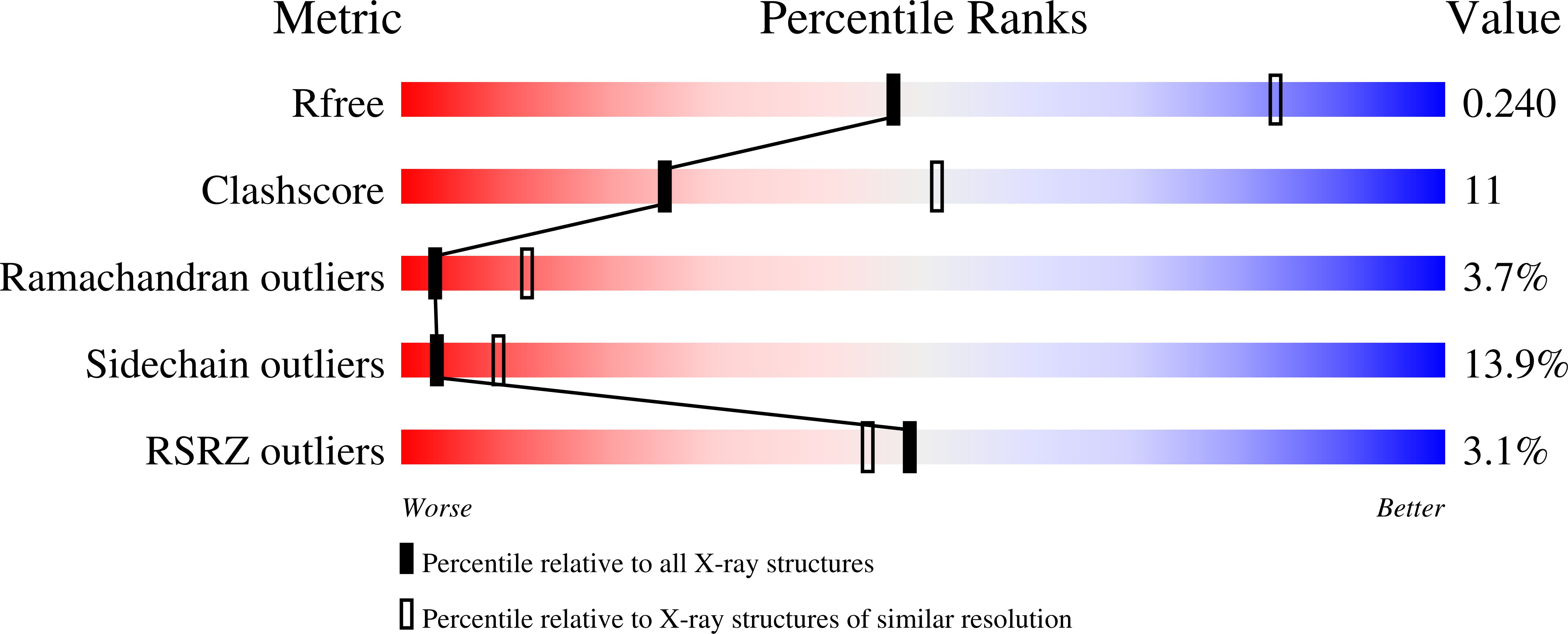
Resolution:
2.89 Å
R-Value Free:
0.24
R-Value Work:
0.21
R-Value Observed:
0.21
Space Group:
I 2 3
Macromolecular Entities
Structures with similar UniProt ID
Protein Blast
Polymer Type:polypeptide(L)
Molecule:Capsid protein
Gene (Uniprot):ORF2
Chain IDs:A, B, C (auth: D), D (auth: E), E (auth: F), F (auth: G), G (auth: H), H (auth: I), I (auth: J), J (auth: K), K (auth: L), L (auth: M), M (auth: N), N (auth: O), O (auth: P)
Chain Length:380
Number of Molecules:15
Biological Source:Cucumber necrosis virus
Primary Citation
Atomic structure of cucumber necrosis virus and the role of the capsid in vector transmission.
J. Virol. 87 12166 12175 (2013)
PMID: 24006433 DOI: 10.1128/JVI.01965-13

Abstact

Cucumber Necrosis Virus (CNV) is a member of the genus Tombusvirus and has a monopartite positive-sense RNA genome packaged in a T=3 icosahedral particle. CNV is transmitted in nature via zoospores of the fungus Olpidium bornovanus. CNV undergoes a conformational change upon binding to the zoospore that is required for transmission, and specific polysaccharides on the zoospore surface have been implicated in binding. To better understand this transmission process, we have determined the atomic structure of CNV. As expected, being a member of the Tombusvirus genus, the core structure of CNV is highly similar to that of Tomato bushy stunt virus (TBSV), with major differences lying on the exposed loops. Also, as was seen with TBSV, CNV appears to have a calcium binding site between the subunits around the quasi-3-fold axes. However, unlike TBSV, there appears to be a novel zinc binding site within the β annulus formed by the N termini of the three C subunits at the icosahedral 3-fold axes. Two of the mutations causing defective transmission map immediately around this zinc binding site. The other mutations causing defective transmission and particle formation are mapped onto the CNV structure, and it is likely that a number of the mutations affect zoospore transmission by affecting conformational transitions rather than directly affecting receptor binding.

Legend

Protein

Chemical

Disease

Primary Citation of related structures
Feedback Form
Name
Email
Institute
Feedback