4D0A image
Deposition Date 2014-04-25
Release Date 2014-12-10
Last Version Date 2023-12-20
Entry Detail
PDB ID:
4D0A
Title:
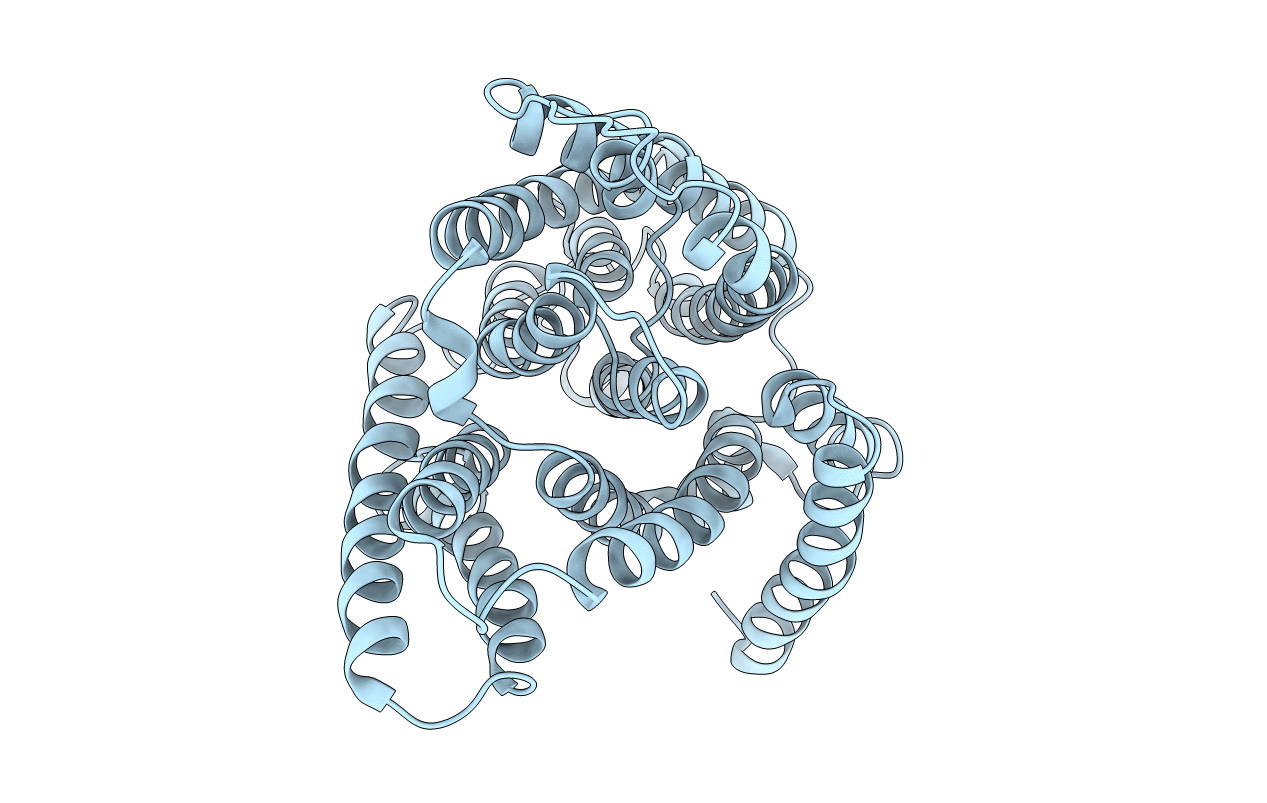
3D EM map of the sodium proton antiporter MjNhaP1 from Methanocaldococcus jannaschii
Biological Source:
Source Organism(s):
Expression System(s):
Method Details:
Experimental Method:
Resolution:
6.00 Å
Space Group:
P 21 21 2
Macromolecular Entities
Polymer Type:polypeptide(L)
Molecule:NA(+)/H(+) ANTIPORTER 1
Gene (Uniprot):MJ0057
Chain IDs:A (auth: B)
Chain Length:426
Number of Molecules:1
Biological Source:METHANOCALDOCOCCUS JANNASCHII DSM 2661
Ligand Molecules
Primary Citation
3D Em Map of the Sodium Proton Antiporter Mjnhap1 from Methanocaldococcus Jannaschii
Elife 3 ? ? (2014)
PMID: 25426803 DOI: 10.7554/ELIFE.03583

Abstact

Sodium/proton antiporters are essential for sodium and pH homeostasis and play a major role in human health and disease. We determined the structures of the archaeal sodium/proton antiporter MjNhaP1 in two complementary states. The inward-open state was obtained by x-ray crystallography in the presence of sodium at pH 8, where the transporter is highly active. The outward-open state was obtained by electron crystallography without sodium at pH 4, where MjNhaP1 is inactive. Comparison of both structures reveals a 7° tilt of the 6 helix bundle. (22)Na(+) uptake measurements indicate non-cooperative transport with an activity maximum at pH 7.5. We conclude that binding of a Na(+) ion from the outside induces helix movements that close the extracellular cavity, open the cytoplasmic funnel, and result in a ∼5 Å vertical relocation of the ion binding site to release the substrate ion into the cytoplasm.

Legend

Protein

Chemical

Disease

Primary Citation of related structures
Feedback Form
Name
Email
Institute
Feedback