Deposition Date
2013-12-05
Release Date
2014-02-26
Last Version Date
2024-11-06
Entry Detail
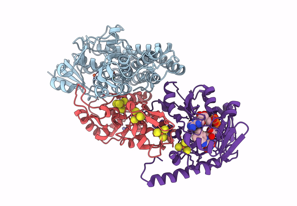
PDB ID:
4CI0
Keywords:
Title:
Electron cryo-microscopy of F420-reducing NiFe hydrogenase Frh
Biological Source:
Source Organism(s):
METHANOTHERMOBACTER MARBURGENSIS (Taxon ID: 145263)
Method Details:
Experimental Method:
Resolution:
3.36 Å
Aggregation State:
PARTICLE
Reconstruction Method:
SINGLE PARTICLE