

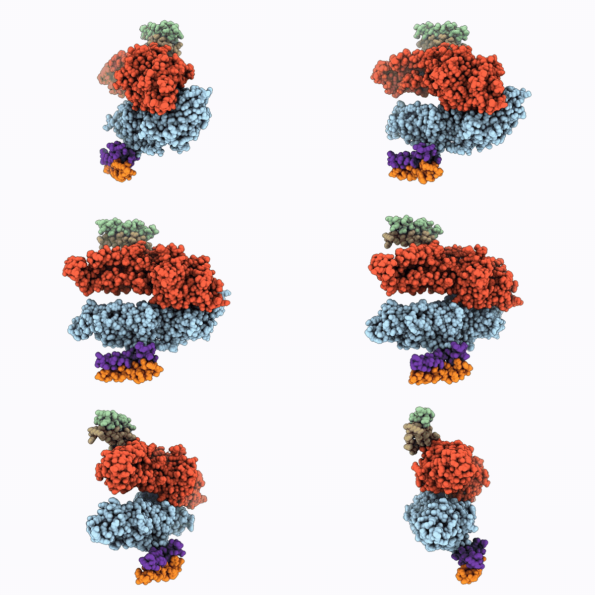
45LQ image
Deposition Date 2026-09-02
Release Date 2026-10-07
Last Version Date 2026-10-14
Entry Detail
PDB ID:
45LQ
Title:
Crystal structure of Escherichia coli YidC in complex with YibN
Biological Source:
Source Organism(s):
Escherichia coli (Taxon ID: 562)
Expression System(s):
Method Details:
Experimental Method:
Resolution:
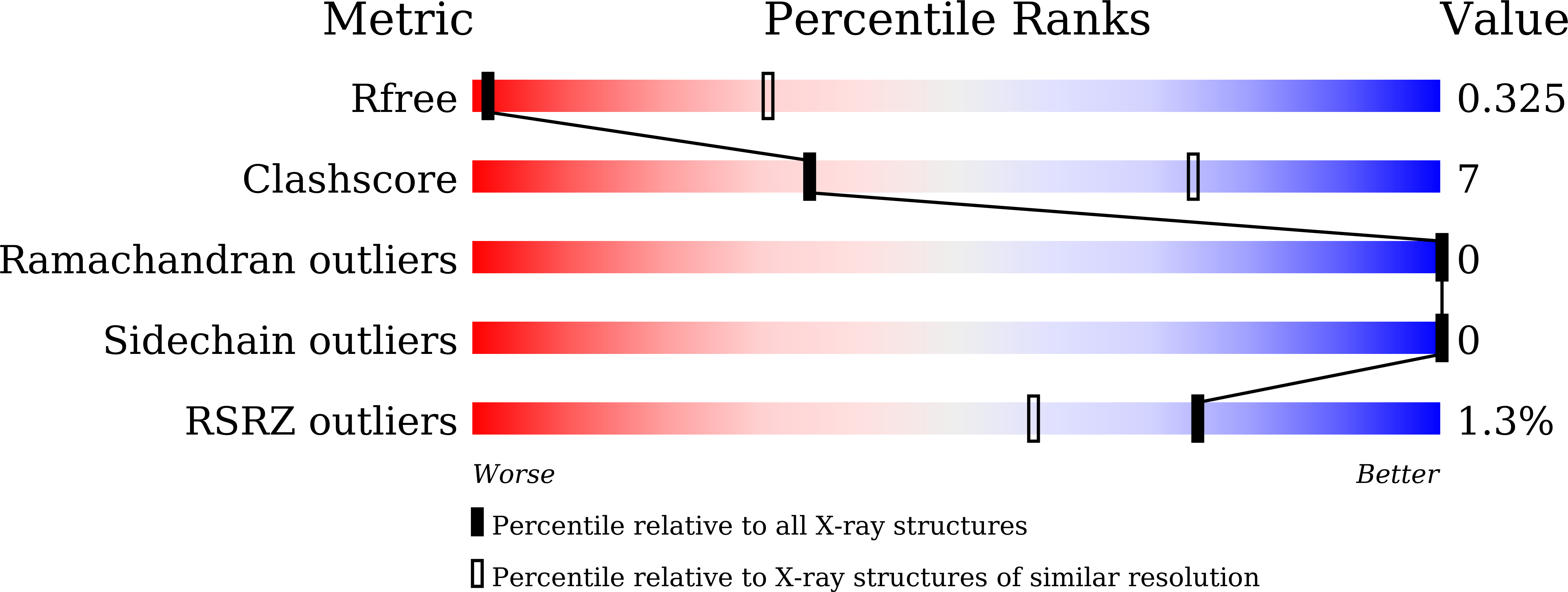
4.09 Å
R-Value Free:
0.32
R-Value Work:
0.26
R-Value Observed:
0.27
Space Group:
P 1 21 1
Macromolecular Entities
Polymer Type:polypeptide(L)
Molecule:Membrane protein insertase Yi
Gene (Uniprot):yidC
Chain IDs:A, B (auth: D)
Chain Length:548
Number of Molecules:2
Biological Source:Escherichia coli
Polymer Type:polypeptide(L)
Molecule:Membrane protein insertase Yi
Gene (Uniprot):yidC
Chain IDs:C (auth: B), E
Chain Length:27
Number of Molecules:2
Biological Source:Escherichia coli
Polymer Type:polypeptide(L)
Molecule:YibN
Gene (Uniprot):yibN
Chain IDs:D (auth: C), F
Chain Length:167
Number of Molecules:2
Biological Source:Escherichia coli
Ligand Molecules
Primary Citation
Crystal structure of YidC in complex with YibN.
Biochem.Biophys.Res.Commun. 838 154668 154668 (2026)
PMID: 42815374 DOI: 10.1016/j.bbrc.2026.154668

Abstact

YidC is a widely conserved membrane protein that facilitates membrane protein insertion and folding. Escherichia coli YidC contains a conserved core region with five transmembrane helices and an additional N-terminal transmembrane helix, TM1. TM1 has not been resolved in previously reported E. coli YidC structures, leaving its position and structural role unclear. YibN is a single-pass inner membrane protein known to interact with YidC, but the structural basis of their association remains unknown. Here, we determined the crystal structure of the E. coli YidC-YibN complex. The structure reveals that TM1 is positioned away from the YidC core and forms the major intramembrane interface with the N-terminal amphipathic helix and transmembrane helix of YibN. YibN also makes additional contacts with the YidC core. Comparison with previously reported YidC structures further shows that the paddle domain is positioned closer to TM5 while retaining its overall fold. These findings define a previously unresolved role of YidC TM1 in YibN association and suggest that YibN binding may be coupled to repositioning of the YidC paddle domain.

Legend

Protein

Chemical

Disease

Primary Citation of related structures
Feedback Form
Name
Email
Institute
Feedback