

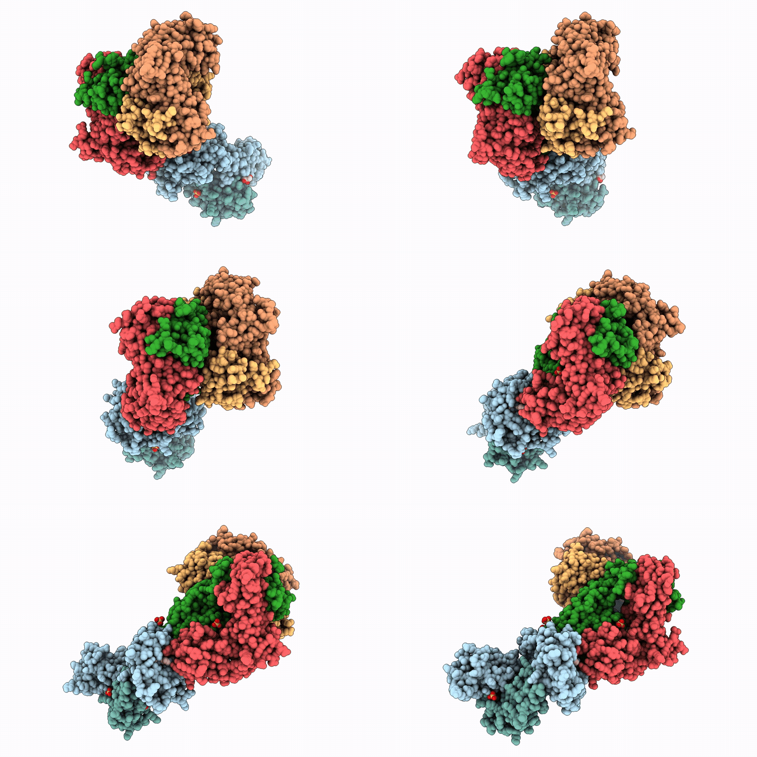
43RH image
Deposition Date 2026-07-14
Release Date 2026-07-29
Last Version Date 2026-07-29
Entry Detail
PDB ID:
43RH
Keywords:
Title:
Crystal structure of TCR C3K in complex with H2-Q9/VP2.139
Biological Source:
Source Organism(s):
Expression System(s):
Method Details:
Experimental Method:
Resolution:
3.33 Å
R-Value Free:
0.26
R-Value Work:
0.21
R-Value Observed:
0.22
Space Group:
C 1 2 1

Macromolecular Entities
Polymer Type:polypeptide(L)
Molecule:Histocompatibility 2, Q regio
Gene (Uniprot):H2-Q9
Chain IDs:A
Chain Length:274
Number of Molecules:1
Biological Source:Mus musculus
Polymer Type:polypeptide(L)
Molecule:Beta-2-microglobulin
Gene (Uniprot):B2m
Chain IDs:B
Chain Length:99
Number of Molecules:1
Biological Source:Mus musculus
Structural Superimposition Protein Blast
Polymer Type:polypeptide(L)
Molecule:T cell receptor alpha variabl
Gene (Uniprot):Trav13d-1, Traj18, TRAC
Chain IDs:C, F (auth: E)
Chain Length:201
Number of Molecules:2
Biological Source:Mus musculus
Structural Superimposition Protein Blast
Polymer Type:polypeptide(L)
Molecule:T cell receptor beta, variabl
Gene (Uniprot):Trbj1-1, Trbv2, TRBC1
Chain IDs:D, G (auth: F)
Chain Length:241
Number of Molecules:2
Biological Source:Mus musculus
Polymer Type:polypeptide(L)
Molecule:Minor capsid protein VP2
Chain IDs:E (auth: P)
Chain Length:9
Number of Molecules:1
Biological Source:Alphapolyomavirus muris
Primary Citation
A murine MHC-Ib molecule, H2-Q9, drives peptide-centric T cell receptor recognition of a viral antigen
To Be Published ? ? ? (?)
Primary Citation of related structures
Feedback Form
Name
Email
Institute
Feedback