

3WSQ image
Deposition Date 2014-03-20
Release Date 2015-03-25
Last Version Date 2024-11-13
Entry Detail
PDB ID:
3WSQ
Title:
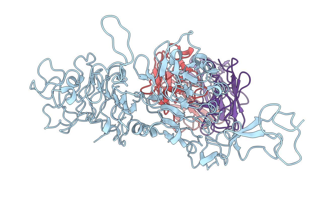
Structure of HER2 with an Fab
Biological Source:
Source Organism(s):
Homo sapiens (Taxon ID: 9606)
Expression System(s):
Method Details:
Experimental Method:
Resolution:
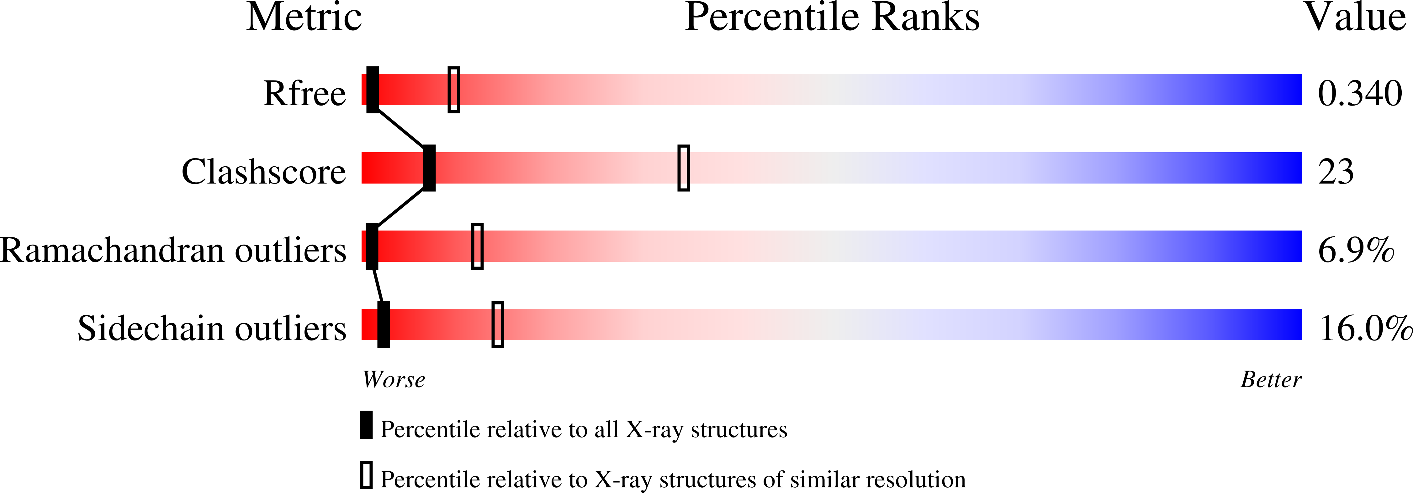
3.50 Å
R-Value Free:
0.25
R-Value Work:
0.22
R-Value Observed:
0.22
Space Group:
P 1 21 1
Macromolecular Entities
Structures with similar UniProt ID
Protein Blast
Polymer Type:polypeptide(L)
Molecule:Receptor tyrosine-protein kin
Gene (Uniprot):ERBB2
Chain IDs:A
Chain Length:564
Number of Molecules:1
Biological Source:Homo sapiens
Protein Blast
Polymer Type:polypeptide(L)
Molecule:Antibody Heavy Chain
Chain IDs:C (auth: H)
Chain Length:218
Number of Molecules:1
Biological Source:Homo sapiens
Protein Blast
Polymer Type:polypeptide(L)
Molecule:Antibody Light Chain
Chain IDs:B (auth: L)
Chain Length:215
Number of Molecules:1
Biological Source:Homo sapiens
Ligand Molecules
Primary Citation
Insights into HER2 signaling from step-by-step optimization of anti-HER2 antibodies.
MAbs 6 978 990 (2014)
PMID: 24838231 DOI: 10.4161/mabs.28786

Abstact

HER2, a ligand-free tyrosine kinase receptor of the HER family, is frequently overexpressed in breast cancer. The anti-HER2 antibody trastuzumab has shown significant clinical benefits in metastatic breast cancer; however, resistance to trastuzumab is common. The development of monoclonal antibodies that have complementary mechanisms of action results in a more comprehensive blockade of ErbB2 signaling, especially HER2/HER3 signaling. Use of such antibodies may have clinical benefits if these antibodies can become widely accepted. Here, we describe a novel anti-HER2 antibody, hHERmAb-F0178C1, which was isolated from a screen of a phage display library. A step-by-step optimization method was employed to maximize the inhibitory effect of this anti-HER2 antibody. Crystallographic analysis was used to determine the three-dimensional structure to 3.5 Å resolution, confirming that the epitope of this antibody is in domain III of HER2. Moreover, this novel anti-HER2 antibody exhibits superior efficacy in blocking HER2/HER3 heterodimerization and signaling, and its use in combination with pertuzumab has a synergistic effect. Characterization of this antibody revealed the important role of a ligand binding site within domain III of HER2. The results of this study clearly indicate the unique potential of hHERmAb-F0178C1, and its complementary inhibition effect on HER2/HER3 signaling warrants its consideration as a promising clinical treatment.

Legend

Protein

Chemical

Disease

Primary Citation of related structures
Feedback Form
Name
Email
Institute
Feedback