

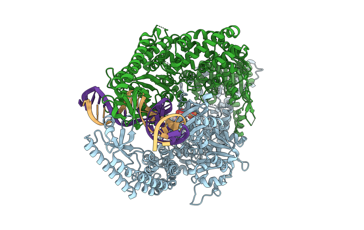
3THY image
Deposition Date 2011-08-19
Release Date 2011-12-21
Last Version Date 2024-02-28
Entry Detail
PDB ID:
3THY
Title:
Human MutSbeta complexed with an IDL of 2 bases (Loop2) and ADP
Biological Source:
Source Organism(s):
Homo sapiens (Taxon ID: 9606)
Expression System(s):
Method Details:
Experimental Method:
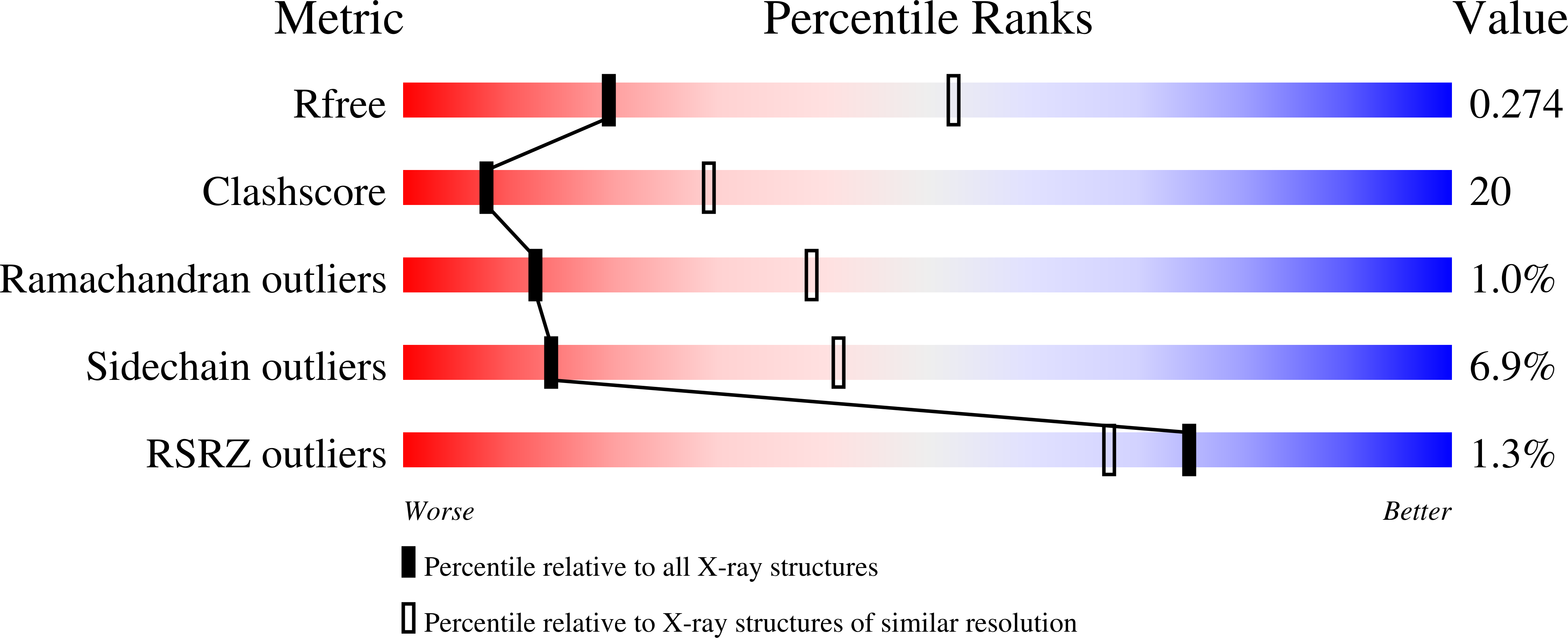
Resolution:
2.89 Å
R-Value Free:
0.27
R-Value Work:
0.19
R-Value Observed:
0.19
Space Group:
P 1
Macromolecular Entities
Polymer Type:polypeptide(L)
Molecule:DNA mismatch repair protein M
Gene (Uniprot):MSH2
Chain IDs:A
Chain Length:934
Number of Molecules:1
Biological Source:Homo sapiens
Polymer Type:polypeptide(L)
Molecule:DNA mismatch repair protein M
Chain IDs:B
Chain Length:918
Number of Molecules:1
Biological Source:Homo sapiens
Polymer Type:polydeoxyribonucleotide
Molecule:DNA Loop2 minus strand
Chain IDs:C (auth: D)
Chain Length:24
Number of Molecules:1
Biological Source:
Polymer Type:polydeoxyribonucleotide
Molecule:DNA Loop2 plus strand
Chain IDs:D (auth: E)
Chain Length:24
Number of Molecules:1
Biological Source:
Ligand Molecules
Primary Citation
Mechanism of mismatch repair revealed by human MutSbeta bound to unpaired DNA loops
Nat. Struct. Mol. Biol. 19 72 78 (2012)
PMID: 22179786 DOI: 10.1038/nsmb.2175

Abstact

DNA mismatch repair corrects replication errors, thus reducing mutation rates and microsatellite instability. Genetic defects in this pathway cause Lynch syndrome and various cancers in humans. Binding of a mispaired or unpaired base by bacterial MutS and eukaryotic MutSα is well characterized. We report here crystal structures of human MutSβ in complex with DNA containing insertion-deletion loops (IDL) of two, three, four or six unpaired nucleotides. In contrast to eukaryotic MutSα and bacterial MutS, which bind the base of a mismatched nucleotide, MutSβ binds three phosphates in an IDL. DNA is severely bent at the IDL; unpaired bases are flipped out into the major groove and partially exposed to solvent. A normal downstream base pair can become unpaired; a single unpaired base can thereby be converted to an IDL of two nucleotides and recognized by MutSβ. The C-terminal dimerization domains form an integral part of the MutS structure and coordinate asymmetrical ATP hydrolysis by Msh2 and Msh3 with mismatch binding to signal for repair.

Legend

Protein

Chemical

Disease

Primary Citation of related structures
Feedback Form
Name
Email
Institute
Feedback