

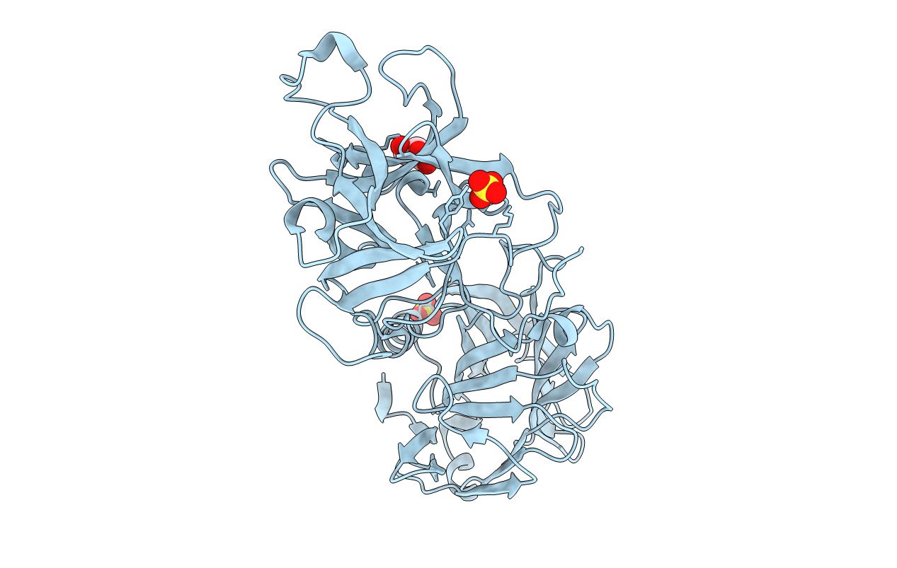
3PME image
Deposition Date 2010-11-16
Release Date 2010-12-15
Last Version Date 2024-11-27
Entry Detail
PDB ID:
3PME
Keywords:
Title:
Crystal structure of the receptor binding domain of botulinum neurotoxin C/D mosaic serotype
Biological Source:
Source Organism(s):
Expression System(s):
Method Details:
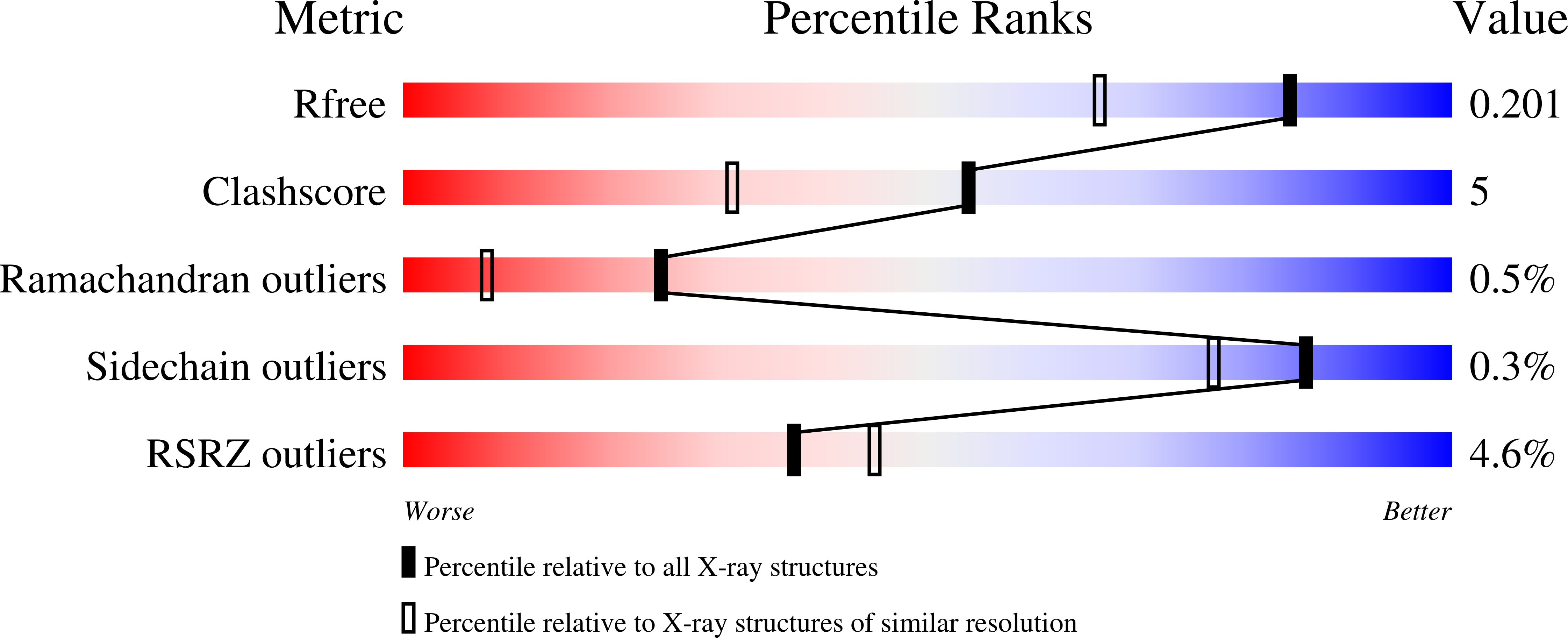
Experimental Method:
Resolution:
1.56 Å
R-Value Free:
0.20
R-Value Work:
0.17
R-Value Observed:
0.17
Space Group:
P 1 21 1
Macromolecular Entities
Polymer Type:polypeptide(L)
Molecule:Type C neurotoxin
Gene (Uniprot):C
Chain IDs:A
Chain Length:420
Number of Molecules:1
Biological Source:Clostridium botulinum
Primary Citation
Crystal structure of the receptor binding domain of the botulinum C-D mosaic neurotoxin reveals potential roles of lysines 1118 and 1136 in membrane interactions.
Biochem.Biophys.Res.Commun. 404 407 412 (2011)
PMID: 21130733 DOI: 10.1016/j.bbrc.2010.11.134

Abstact

The botulinum neurotoxins (BoNTs) produced by different strains of the bacterium Clostridium botulinum are responsible for the disease botulism and include a group of immunologically distinct serotypes (A, B, E, and F) that are considered to be the most lethal natural proteins known for humans. Two BoNT serotypes, C and D, while rarely associated with human infection, are responsible for deadly botulism outbreaks afflicting animals. Also associated with animal infections is the BoNT C-D mosaic protein (BoNT/CD), a BoNT subtype that is essentially a hybrid of the BoNT/C (∼two-third) and BoNT/D (∼one-third) serotypes. While the amino acid sequence of the heavy chain receptor binding (HCR) domain of BoNT/CD (BoNT/CD-HCR) is very similar to the corresponding amino acid sequence of BoNT/D, BoNT/CD-HCR binds synaptosome membranes better than BoNT/D-HCR. To obtain structural insights for the different membrane binding properties, the crystal structure of BoNT/CD-HCR (S867-E1280) was determined at 1.56 Å resolution and compared to previously reported structures for BoNT/D-HCR. Overall, the BoNT/CD-HCR structure is similar to the two sub-domain organization observed for other BoNT HCRs: an N-terminal jellyroll barrel motif and a C-terminal β-trefoil fold. Comparison of the structure of BoNT/CD-HCR with BoNT/D-HCR indicates that K1118 has a similar structural role as the equivalent residue, E1114, in BoNT/D-HCR, while K1136 has a structurally different role than the equivalent residue, G1132, in BoNT/D-HCR. Lysine-1118 forms a salt bridge with E1247 and may enhance membrane interactions by stabilizing the putative membrane binding loop (K1240-N1248). Lysine-1136 is observed on the surface of the protein. A sulfate ion bound to K1136 may mimic a natural interaction with the negatively changed phospholipid membrane surface. Liposome-binding experiments demonstrate that BoNT/CD-HCR binds phosphatidylethanolamine liposomes more tightly than BoNT/D-HCR.

Legend

Protein

Chemical

Disease

Primary Citation of related structures
Feedback Form
Name
Email
Institute
Feedback