

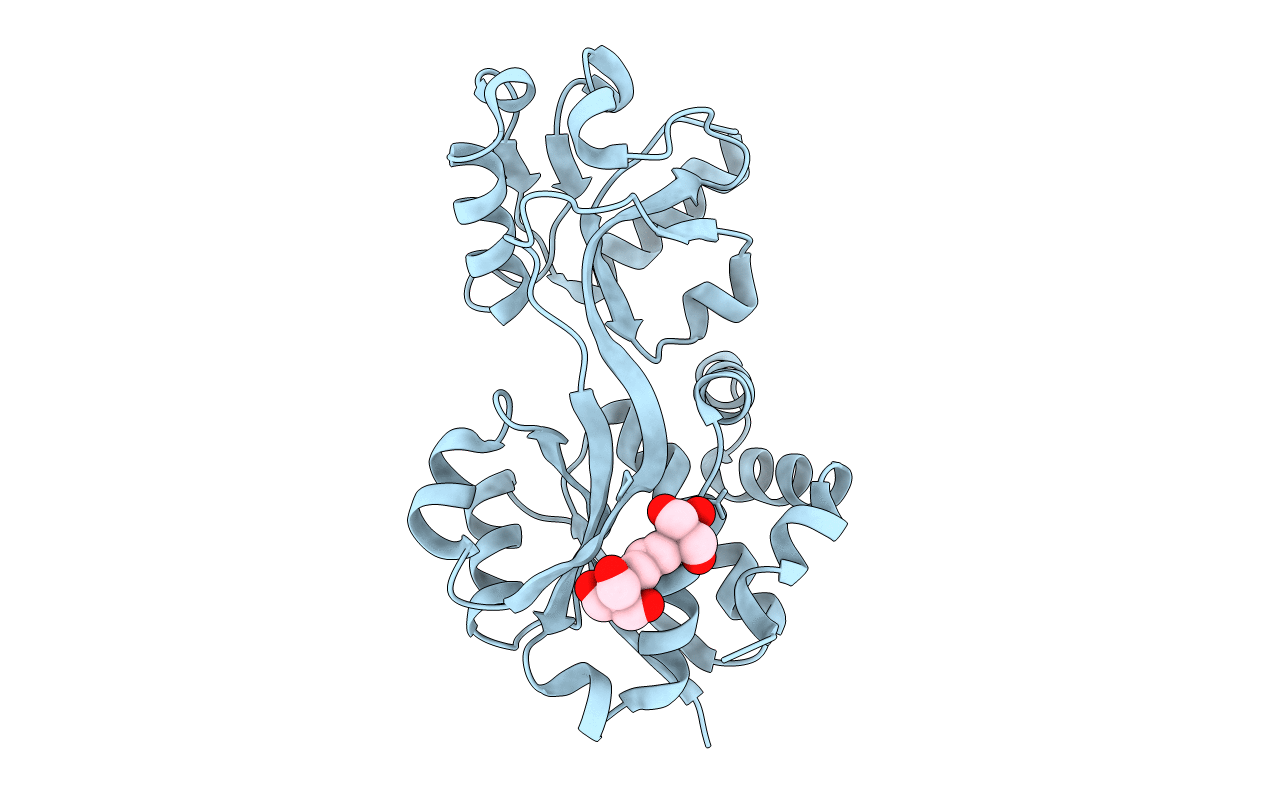
3L6G image
Deposition Date 2009-12-23
Release Date 2010-05-19
Last Version Date 2023-09-06
Entry Detail
PDB ID:
3L6G
Title:
Crystal structure of lactococcal OpuAC in its open conformation
Biological Source:
Source Organism(s):
Expression System(s):
Method Details:
Experimental Method:
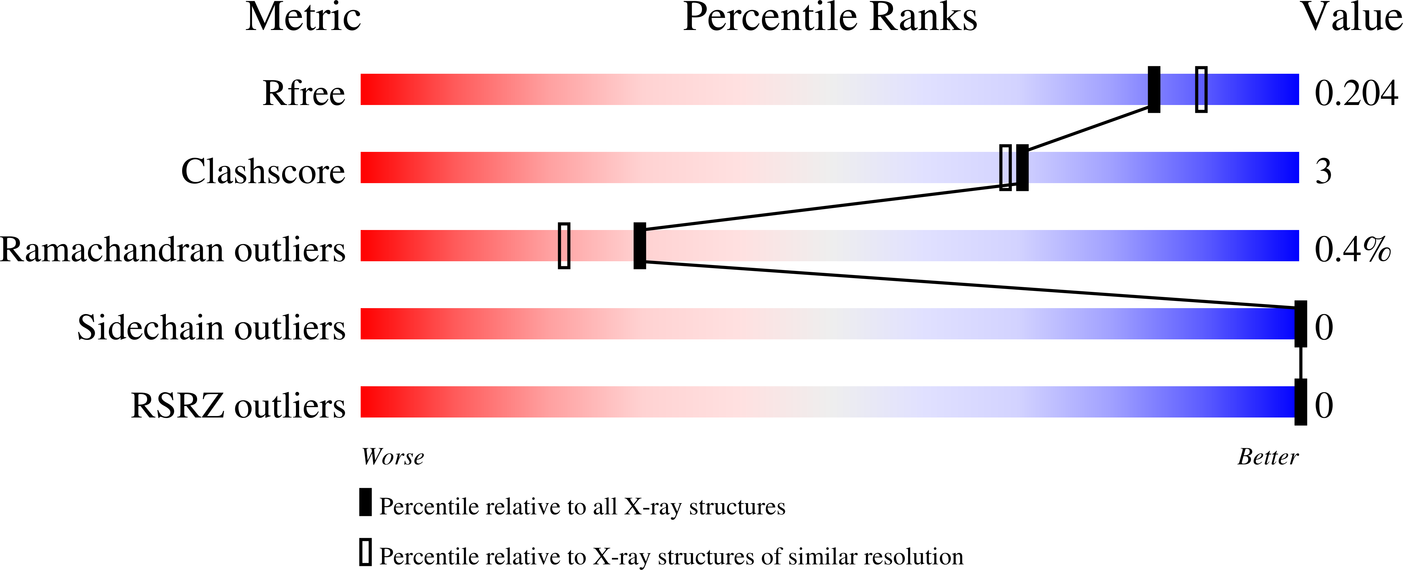
Resolution:
1.90 Å
R-Value Free:
0.20
R-Value Work:
0.16
R-Value Observed:
0.16
Space Group:
P 41 21 2
Macromolecular Entities
Polymer Type:polypeptide(L)
Molecule:Betaine ABC transporter perme
Gene (Uniprot):busAB
Chain IDs:A
Chain Length:256
Number of Molecules:1
Biological Source:Lactococcus lactis
Ligand Molecules
Primary Citation
Ligand binding and crystal structures of the substrate-binding domain of the ABC transporter OpuA.
PLoS ONE 5 e10361 e10361 (2010)
PMID: 20454456 DOI: 10.1371/journal.pone.0010361

Abstact

BACKGROUND The ABC transporter OpuA from Lactococcus lactis transports glycine betaine upon activation by threshold values of ionic strength. In this study, the ligand binding characteristics of purified OpuA in a detergent-solubilized state and of its substrate-binding domain produced as soluble protein (OpuAC) was characterized. PRINCIPAL FINDINGS The binding of glycine betaine to purified OpuA and OpuAC (K(D) = 4-6 microM) did not show any salt dependence or cooperative effects, in contrast to the transport activity. OpuAC is highly specific for glycine betaine and the related proline betaine. Other compatible solutes like proline and carnitine bound with affinities that were 3 to 4 orders of magnitude lower. The low affinity substrates were not noticeably transported by membrane-reconstituted OpuA. OpuAC was crystallized in an open (1.9 A) and closed-liganded (2.3 A) conformation. The binding pocket is formed by three tryptophans (Trp-prism) coordinating the quaternary ammonium group of glycine betaine in the closed-liganded structure. Even though the binding site of OpuAC is identical to that of its B. subtilis homolog, the affinity for glycine betaine is 4-fold higher. CONCLUSIONS Ionic strength did not affect substrate binding to OpuA, indicating that regulation of transport is not at the level of substrate binding, but rather at the level of translocation. The overlap between the crystal structures of OpuAC from L.lactis and B.subtilis, comprising the classical Trp-prism, show that the differences observed in the binding affinities originate from outside of the ligand binding site.

Legend

Protein

Chemical

Disease

Primary Citation of related structures
Feedback Form
Name
Email
Institute
Feedback