

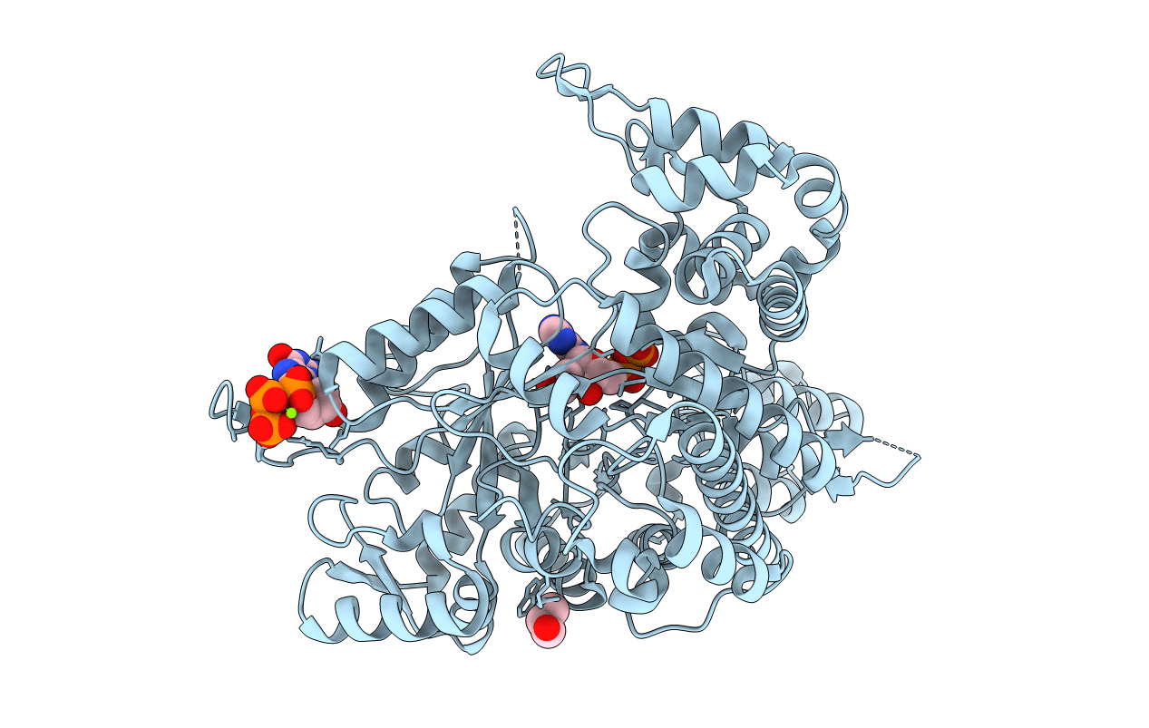
3K8T image
Deposition Date 2009-10-14
Release Date 2010-03-02
Last Version Date 2023-09-06
Entry Detail
PDB ID:
3K8T
Keywords:
Title:
Structure of eukaryotic rnr large subunit R1 complexed with designed adp analog compound
Biological Source:
Source Organism(s):
Expression System(s):
Method Details:
Experimental Method:
Resolution:
2.10 Å
R-Value Free:
0.29
R-Value Work:
0.25
R-Value Observed:
0.25
Space Group:
P 21 21 2

Macromolecular Entities
Structures with similar UniProt ID
Protein Blast
Polymer Type:polypeptide(L)
Molecule:Ribonucleoside-diphosphate re
Gene (Uniprot):RNR1
Chain IDs:A
Chain Length:888
Number of Molecules:1
Biological Source:Saccharomyces cerevisiae
Primary Citation
Structure-Based Design, Synthesis, and Evaluation of 2'-(2-Hydroxyethyl)-2'-deoxyadenosine and the 5'-Diphosphate Derivative as Ribonucleotide Reductase Inhibitors
ChemMedChem 4 1649 1656 (2009)
PMID: 19681093 DOI: 10.1002/cmdc.200900236

Abstact

Analysis of the recently solved X-ray crystal structures of Saccharomyces cerevisiae ribonucleotide reductase I (ScRnr1) in complex with effectors and substrates led to the discovery of a conserved water molecule located at the active site that interacted with the 2'-hydroxy group of the nucleoside ribose. In this study 2'-(2-hydroxyethyl)-2'-deoxyadenosine 1 and the 5'-diphosphate derivative 2 were designed and synthesized to see if the conserved water molecule could be displaced by a hydroxymethylene group, to generate novel RNR inhibitors as potential antitumor agents. Herein we report the synthesis of analogues 1 and 2, and the co-crystal structure of adenosine diphosphate analogue 2 bound to ScRnr1, which shows the conserved water molecule is displaced as hypothesized.

Legend

Protein

Chemical

Disease

Primary Citation of related structures
Feedback Form
Name
Email
Institute
Feedback