3K8K image
Deposition Date 2009-10-14
Release Date 2010-03-02
Last Version Date 2024-11-27
Entry Detail
PDB ID:
3K8K
Title:
Crystal structure of SusG
Biological Source:
Source Organism(s):
Expression System(s):
Method Details:
Experimental Method:
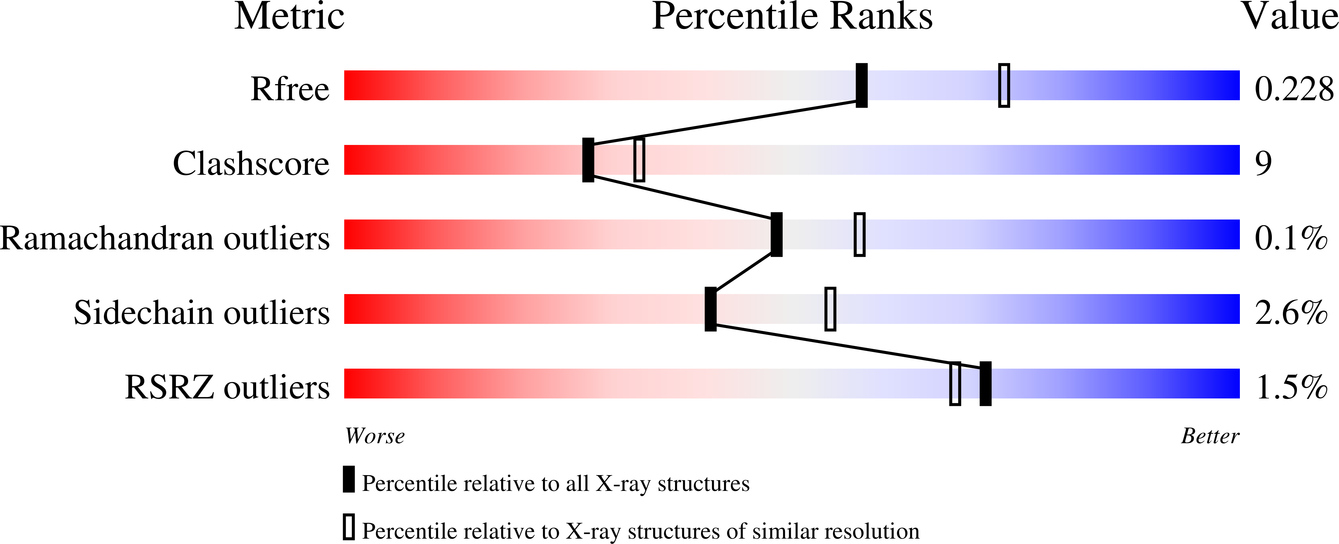
Resolution:
2.20 Å
R-Value Free:
0.23
R-Value Work:
0.19
Space Group:
P 41
Macromolecular Entities
Structures with similar UniProt ID
Protein Blast
Polymer Type:polypeptide(L)
Molecule:Alpha-amylase, susG
Gene (Uniprot):susG
Chain IDs:A, B
Chain Length:669
Number of Molecules:2
Biological Source:Bacteroides thetaiotaomicron
Modified Residue
Compound ID Chain ID Parent Comp ID Details 2D Image
MSE A MET SELENOMETHIONINE
Primary Citation
SusG: A Unique Cell-Membrane-Associated alpha-Amylase from a Prominent Human Gut Symbiont Targets Complex Starch Molecules.
Structure 18 200 215 (2010)
PMID: 20159465 DOI: 10.1016/j.str.2009.12.010

Abstact

SusG is an alpha-amylase and part of a large protein complex on the outer surface of the bacterial cell and plays a major role in carbohydrate acquisition by the animal gut microbiota. Presented here, the atomic structure of SusG has an unusual extended, bilobed structure composed of amylase at one end and an unprecedented internal carbohydrate-binding motif at the other. Structural studies further demonstrate that the carbohydrate-binding motif binds maltooligosaccharide distal to, and on the opposite side of, the amylase catalytic site. SusG has an additional starch-binding site on the amylase domain immediately adjacent to the active cleft. Mutagenesis analysis demonstrates that these two additional starch-binding sites appear to play a role in catabolism of insoluble starch. However, elimination of these sites has only a limited effect, suggesting that they may have a more important role in product exchange with other Sus components.

Legend

Protein

Chemical

Disease

Primary Citation of related structures
Feedback Form
Name
Email
Institute
Feedback