3I3D image
Deposition Date 2009-06-30
Release Date 2010-05-12
Last Version Date 2023-09-06
Entry Detail
PDB ID:
3I3D
Keywords:
Title:
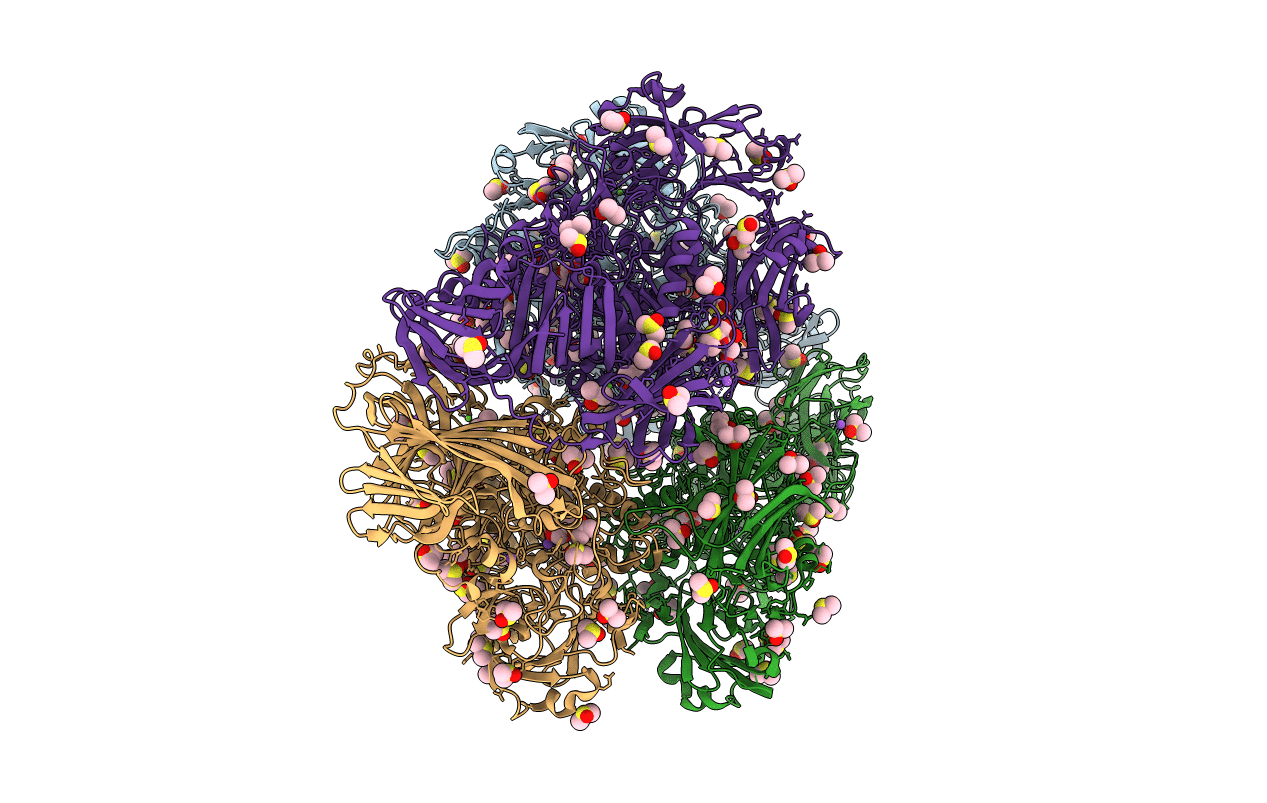
E. COLI (lacZ) BETA-GALACTOSIDASE (M542A) IN COMPLEX WITH IPTG
Biological Source:
Source Organism(s):
Expression System(s):
Method Details:
Experimental Method:
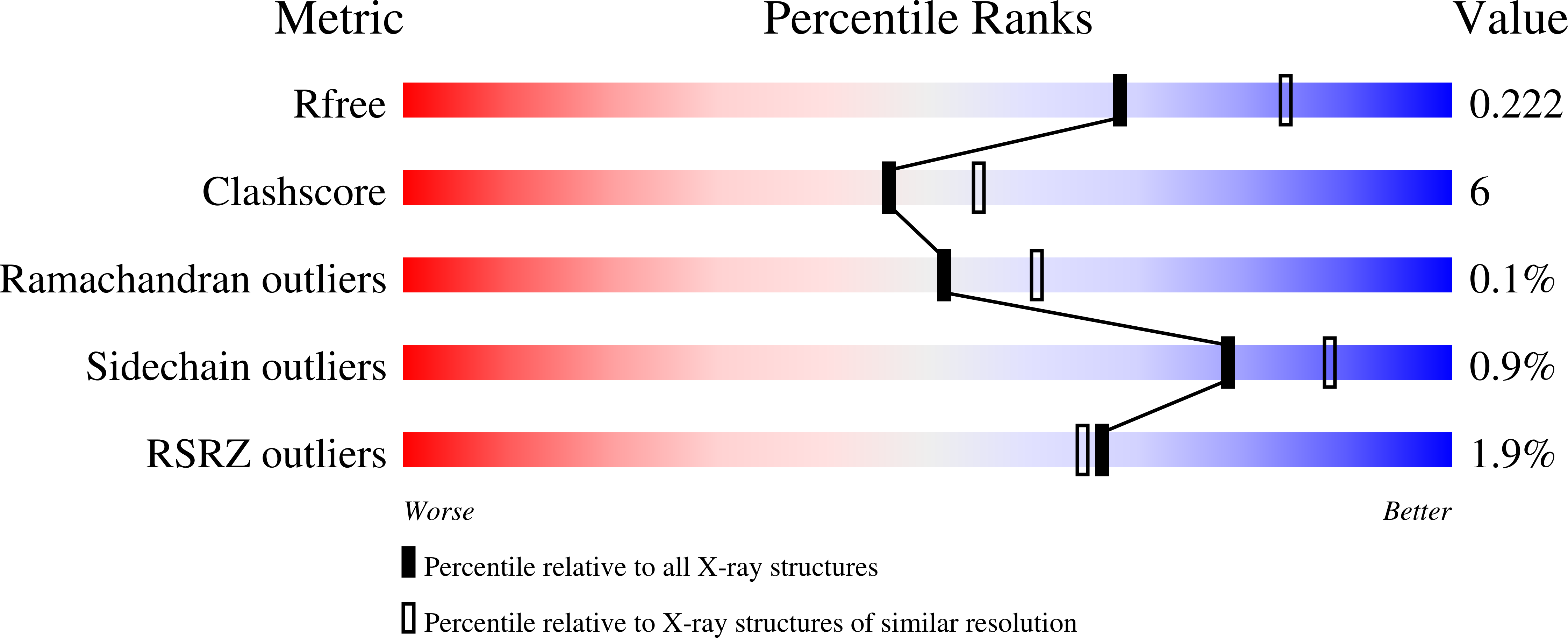
Resolution:
2.20 Å
R-Value Free:
0.23
R-Value Work:
0.18
R-Value Observed:
0.18
Space Group:
P 21 21 21
Macromolecular Entities
Structures with similar UniProt ID
Protein Blast
Polymer Type:polypeptide(L)
Molecule:Beta-galactosidase
Mutagens:M543A
Chain IDs:A, B, C, D
Chain Length:1023
Number of Molecules:4
Biological Source:Escherichia coli
Primary Citation
Role of Met-542 as a guide for the conformational changes of Phe-601 that occur during the reaction of β-galactosidase (Escherichia coli).
Biochem. Cell Biol. 88 861 869 (2010)
PMID: 20921997 DOI: 10.1139/O10-009

Abstact

The Met-542 residue of beta-galactosidase is important for the enzyme's activity because it acts as a guide for the movement of the benzyl side chain of Phe-601 between two stable positions. This movement occurs in concert with an important conformational change (open vs. closed) of an active site loop (residues 794-803). Phe-601 and Arg-599, which interact with each other via the pi electrons of Phe-601 and the guanidium cation of Arg-599, move out of their normal positions and become disordered when Met-542 is replaced by an Ala residue because of the loss of the guide. Since the backbone carbonyl of Phe-601 is a ligand for Na(+), the Na(+) also moves out of its normal position and becomes disordered; the Na(+) binds about 120 times more poorly. In turn, two other Na(+) ligands, Asn-604 and Asp-201, become disordered. A substrate analog (IPTG) restored Arg-599, Phe-601, and Na(+) to their normal open-loop positions, whereas a transition state analog d-galactonolactone) restored them to their normal closed-loop positions. These compounds also restored order to Phe-601, Asn-604, Asp-201, and Na(+). Binding energy was, however, necessary to restore structure and order. The K(s) values of oNPG and pNPG and the competitive K(i) values of substrate analogs were 90-250 times higher than with native enzyme, whereas the competitive K(i) values of transition state analogs were ~3.5-10 times higher. Because of this, the E*S energy level is raised more than the E*transition state energy level and less activation energy is needed for galactosylation. The galactosylation rates (k(2)) of M542A-beta-galactosidase therefore increase. However, the rate of degalactosylation (k(3)) decreased because the E*transition state complex is less stable.

Legend

Protein

Chemical

Disease

Primary Citation of related structures
Feedback Form
Name
Email
Institute
Feedback