

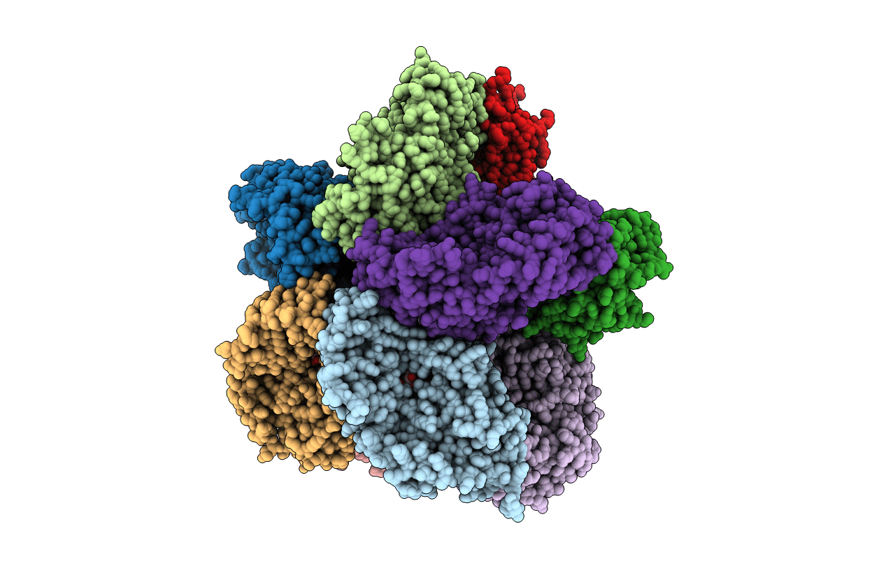
3HDY image
Deposition Date 2009-05-07
Release Date 2009-12-29
Last Version Date 2023-09-06
Entry Detail
PDB ID:
3HDY
Keywords:
Title:
Crystal Structure of UDP-galactopyranose mutase (reduced form) in complex with substrate
Biological Source:
Source Organism(s):
Expression System(s):
Method Details:
Experimental Method:
Resolution:
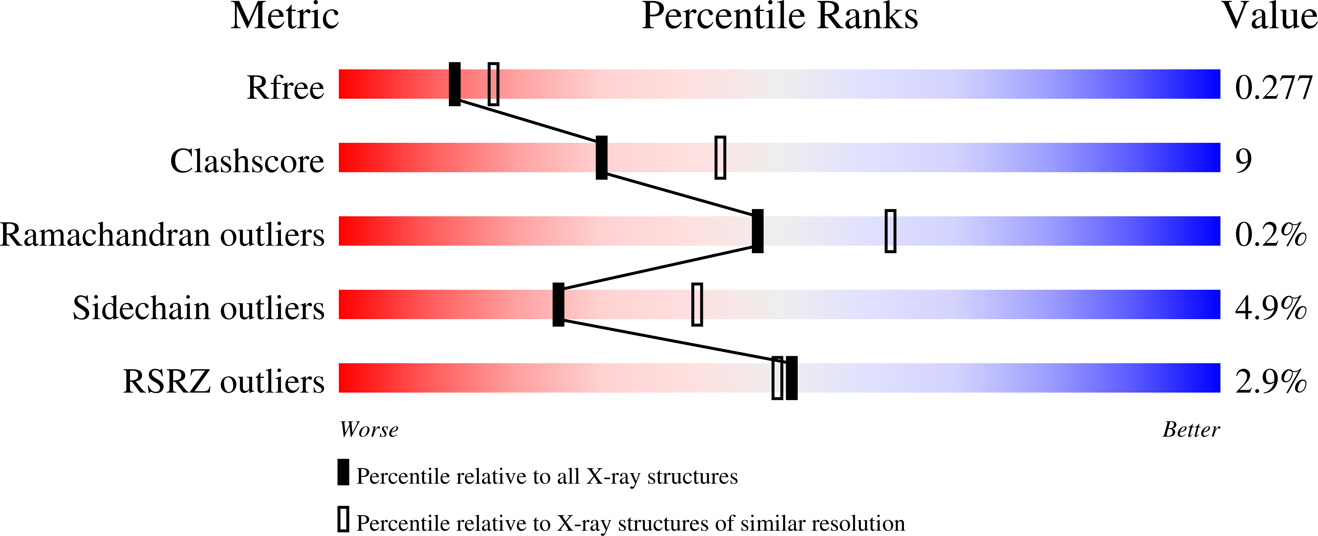
2.40 Å
R-Value Free:
0.28
R-Value Work:
0.23
R-Value Observed:
0.23
Space Group:
P 21 21 21
Macromolecular Entities
Structures with similar UniProt ID
Protein Blast
Polymer Type:polypeptide(L)
Molecule:UDP-galactopyranose mutase
Gene (Uniprot):DR_A0367
Chain IDs:A, B, C, D, E, F, G, H, I, J
Chain Length:397
Number of Molecules:10
Biological Source:Deinococcus radiodurans
Primary Citation
Structural basis of substrate binding to UDP-galactopyranose mutase: crystal structures in the reduced and oxidized state complexed with UDP-galactopyranose and UDP.
J. Mol. Biol. 394 864 877 (2009)
PMID: 19836401 DOI: 10.1016/j.jmb.2009.10.013

Abstact

D-Galactofuranose (Galf) residues are found in the cell walls of pathogenic microbes such as Mycobacterium tuberculosis, and are essential for viability. UDP-galactopyranose mutase (UGM) is a unique flavo-enzyme that catalyzes the reversible conversion of UDP-galactopyranose (UDP-Galp) and UDP-galactofuranose (UDP-Galf). UDP-Galf is the active precursor of Galf residues found in cell walls. Despite the wealth of biochemical/mechanistic data generated for UGM, the structural basis of substrate binding is still lacking. Here, we report the crystal structures of UGM from Deinococcus radiodurans (drUGM) in complex with its natural substrate (UDP-Galp) and UDP. Crystal structures of drUGM:UDP-Galp complexes with oxidized and reduced FAD were determined at 2.36 A and 2.50 A resolution, respectively. The substrate is buried in the active site in an unusual folded conformation and the anomeric carbon of the galactose is at a favorable distance (2.8 A) from N5 of FAD to form an FAD-galactose adduct. The mobile loops in the substrate complex structure exist in a closed conformation. The drUGM-UDP complex structure was determined at 2.55 A resolution and its overall structure is identical with that of the oxidized and reduced complexes, including the conformation of the mobile loops. Comparison with the recently reported UGM:UDP-glucose complex structure reveals key differences and the structures reported here are likely to represent the productive/active conformation of UGM. These structures provide valuable insights into substrate recognition and a basis for understanding the mechanism. These complex structures may serve as a platform for structure-guided design of inhibitors of UGM.

Legend

Protein

Chemical

Disease

Primary Citation of related structures
Feedback Form
Name
Email
Institute
Feedback