

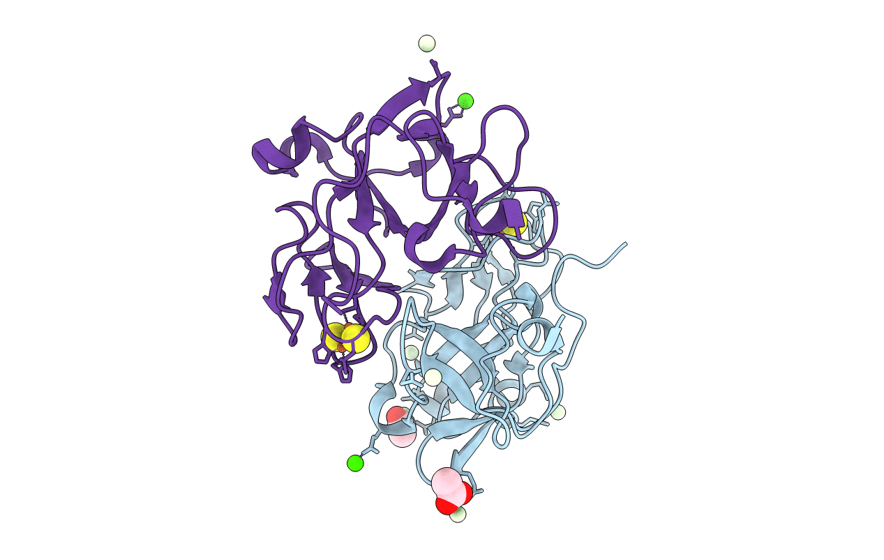
3FOU image
Deposition Date 2009-01-02
Release Date 2009-10-20
Last Version Date 2024-10-30
Entry Detail
PDB ID:
3FOU
Title:
Low pH structure of the Rieske protein from Thermus thermophilus at 2.1 A
Biological Source:
Source Organism(s):
Expression System(s):
Method Details:
Experimental Method:
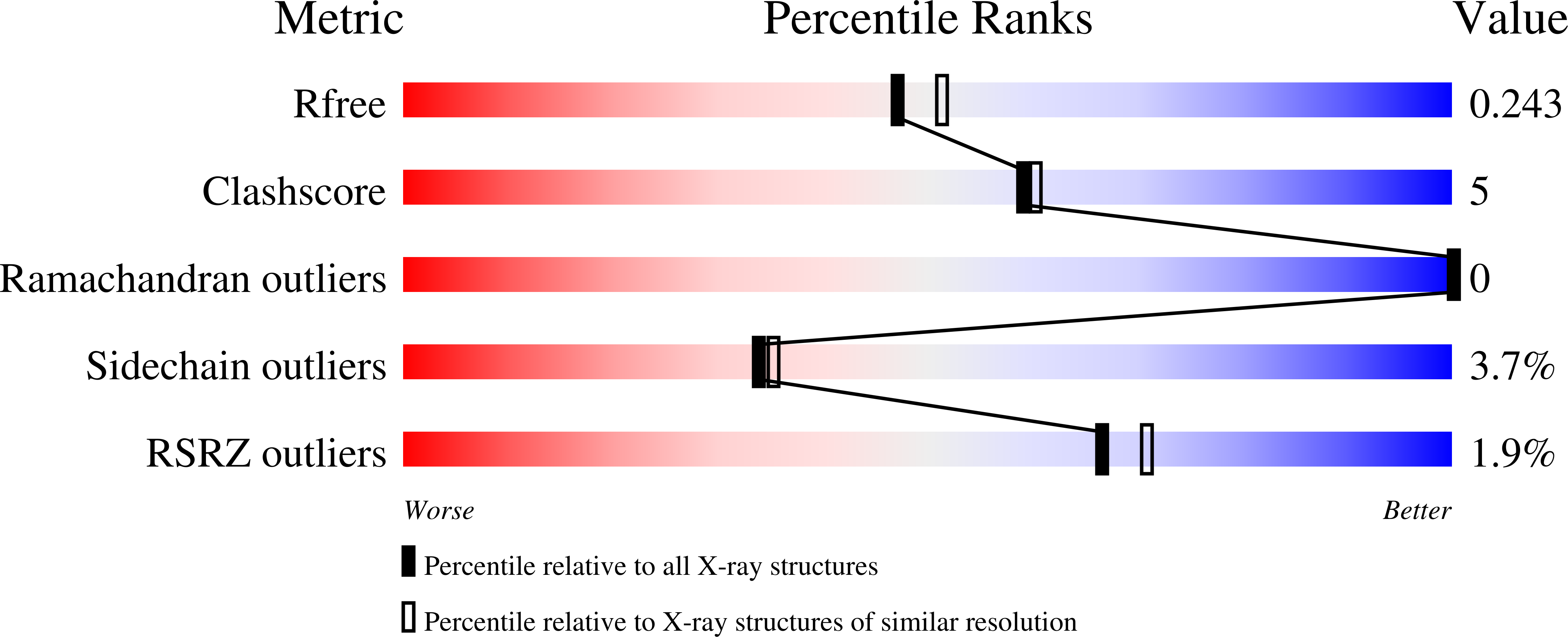
Resolution:
2.10 Å
R-Value Free:
0.24
R-Value Work:
0.19
R-Value Observed:
0.20
Space Group:
P 21 21 2
Macromolecular Entities
Structures with similar UniProt ID
Protein Blast
Polymer Type:polypeptide(L)
Molecule:Quinol-cytochrome c reductase
Gene (Uniprot):TTHA1931
Chain IDs:A, B
Chain Length:156
Number of Molecules:2
Biological Source:Thermus thermophilus
Primary Citation
Effects of pH on the Rieske Protein from Thermus thermophilus: A Spectroscopic and Structural Analysis
Biochemistry 48 9848 9857 (2009)
PMID: 19772300 DOI: 10.1021/bi901126u

Abstact

The Rieske protein from Thermus thermophilus (TtRp) and a truncated version of the protein (truncTtRp), produced to achieve a low-pH crystallization condition, have been characterized using UV-visible and circular dichroism spectroscopies. TtRp and truncTtRp undergo a change in the UV-visible spectra with increasing pH. The LMCT band at 458 nm shifts to 436 nm and increases in intensity. The increase at 436 nm versus pH can be fit using the sum of two Henderson-Hasselbalch equations, yielding two pK(a) values for the oxidized protein. For TtRp, pK(ox1) = 7.48 +/- 0.12 and pK(ox2) = 10.07 +/- 0.17. For truncTtRp, pK(ox1) = 7.87 +/- 0.17 and pK(ox2) = 9.84 +/- 0.42. The shift to shorter wavelength and the increase in intensity for the LMCT band with increasing pH are consistent with deprotonation of the histidine ligands. A pH titration of truncTtRp monitored by circular dichroism also showed pH-dependent changes at 315 and 340 nm. At 340 nm, the fit gives pK(ox1) = 7.14 +/- 0.26 and pK(ox2) = 9.32 +/- 0.36. The change at 315 nm is best fit for a single deprotonation event, giving pK(ox1) = 7.82 +/- 0.10. The lower wavelength region of the CD spectra was unaffected by pH, indicating that the overall fold of the protein remains unchanged, which is consistent with crystallographic results of truncTtRp. The structure of truncTtRp crystallized at pH 6.2 is very similar to TtRp at pH 8.5 and contains only subtle changes localized at the [2Fe-2S] cluster. These titration and structural results further elucidate the histidine ligand characteristics and are consistent with important roles for these amino acids.

Legend

Protein

Chemical

Disease

Primary Citation of related structures
Feedback Form
Name
Email
Institute
Feedback