

3FHP image
Deposition Date 2008-12-09
Release Date 2009-10-20
Last Version Date 2024-11-20
Entry Detail
PDB ID:
3FHP
Keywords:
Title:
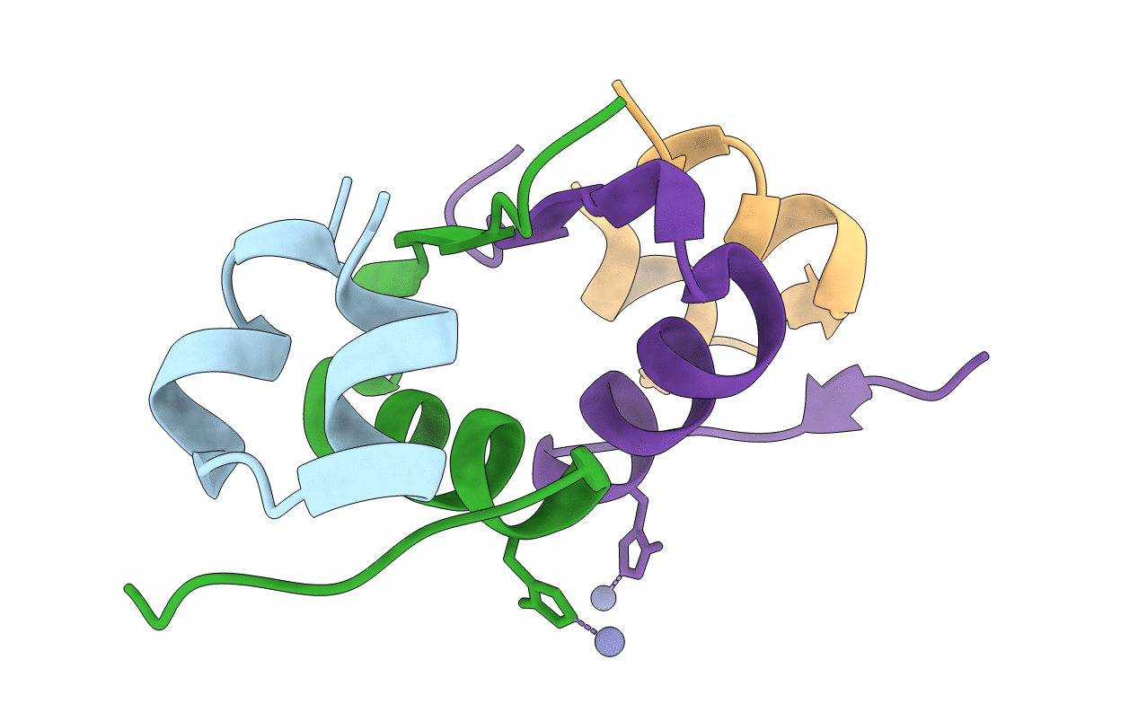
A neutron crystallographic analysis of a porcine 2Zn insulin at 2.0 A resolution
Biological Source:
Source Organism(s):
Sus scrofa (Taxon ID: 9823)
Expression System(s):
Method Details:
Experimental Method:
Resolution:
2.00 Å
R-Value Free:
0.24
R-Value Work:
0.16
R-Value Observed:
0.16
Space Group:
H 3
Macromolecular Entities
Structures with similar UniProt ID
Protein Blast
Polymer Type:polypeptide(L)
Molecule:Insulin
Gene (Uniprot):INS
Chain IDs:A, C
Chain Length:21
Number of Molecules:2
Biological Source:Sus scrofa
Structures with similar UniProt ID
Protein Blast
Polymer Type:polypeptide(L)
Molecule:Insulin
Gene (Uniprot):INS
Chain IDs:B, D
Chain Length:30
Number of Molecules:2
Biological Source:Sus scrofa
Primary Citation
A neutron crystallographic analysis of T6 porcine insulin at 2.1 A resolution
Acta Crystallogr. D Biol. Crystallogr. 65 1042 1050 (2009)
PMID: 19770501 DOI: 10.1107/S090744490902770X

Abstact

Neutron diffraction data for T(6) porcine insulin were collected to 2.1 A resolution from a single crystal partly deuterated by exchange of mother liquor. A maximum-likelihood structure refinement was undertaken using the neutron data and the structure was refined to a residual of 0.179. The hydrogen-bonding network of the central core of the hexamer was observed and the charge balance between positively charged Zn ions and their surrounding structure was interpreted by considering the protonation and/or deprotonation states and interactions of HisB10, water and GluB13. The observed double conformation of GluB13 was essential to interpreting the charge balance and could be compared with the structure of a dried crystal of T(6) human insulin at 100 K. Differences in the dynamic behaviour of the water molecules coordinating the upper and lower Zn ions were observed and interpreted. The hydrogen bonds in the insulin molecules, as well as those involving HisB10 and GluB13, are discussed. The hydrogen/deuterium (H/D) exchange ratios of the amide H atoms of T(6) porcine insulin in crystals were obtained and showed that regions highly protected from H/D exchange are concentrated in the centre of a helical region of the B chains. From the viewpoint of soaking time versus H/D-exchange ratios, the amide H atoms can be classified into three categories.

Legend

Protein

Chemical

Disease

Primary Citation of related structures
Feedback Form
Name
Email
Institute
Feedback