Deposition Date
2008-08-01
Release Date
2009-07-21
Last Version Date
2023-11-01
Entry Detail
PDB ID:
3E17
Keywords:
Title:
Crystal structure of the second PDZ domain from human Zona Occludens-2
Biological Source:
Source Organism(s):
Homo sapiens (Taxon ID: 9606)
Expression System(s):
Method Details:
Experimental Method:
Resolution:
1.75 Å
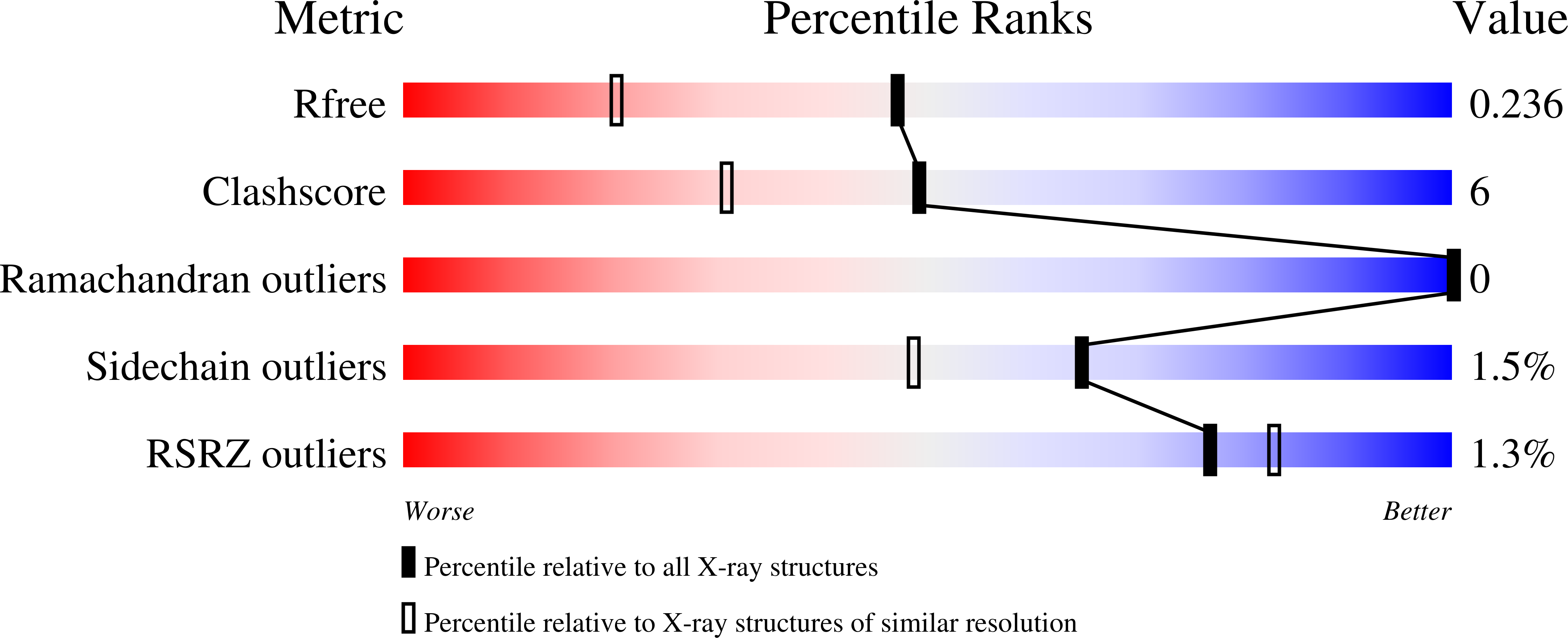
R-Value Free:
0.23
R-Value Work:
0.20
R-Value Observed:
0.20
Space Group:
P 1