

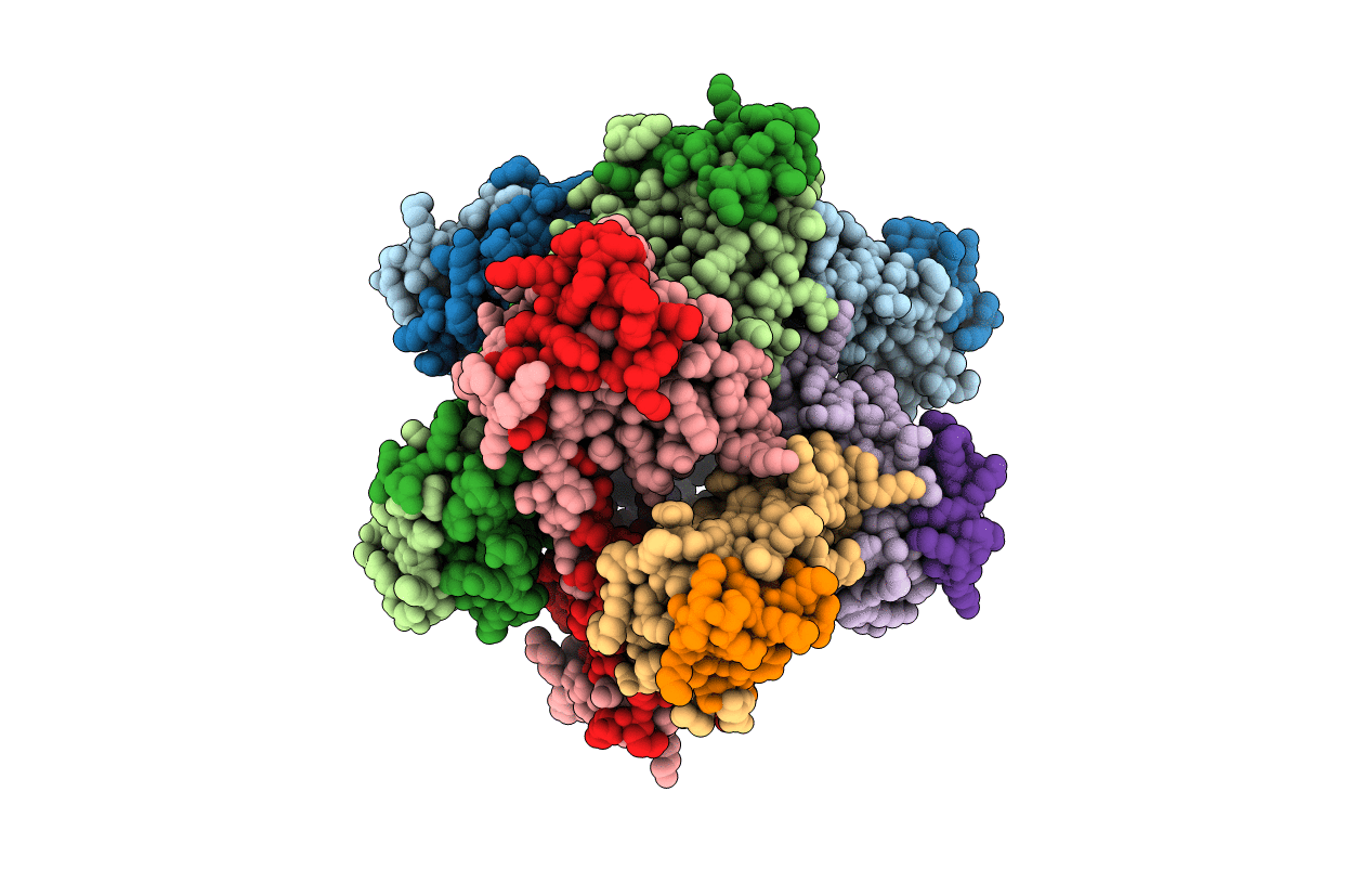
3CAE image
Deposition Date 2008-02-19
Release Date 2008-07-22
Last Version Date 2023-08-30
Entry Detail
PDB ID:
3CAE
Keywords:
Title:
Structure of NNQQNY as an insert in T7 endonuclease I
Biological Source:
Source Organism(s):
Expression System(s):
Method Details:
Experimental Method:
Resolution:
3.00 Å
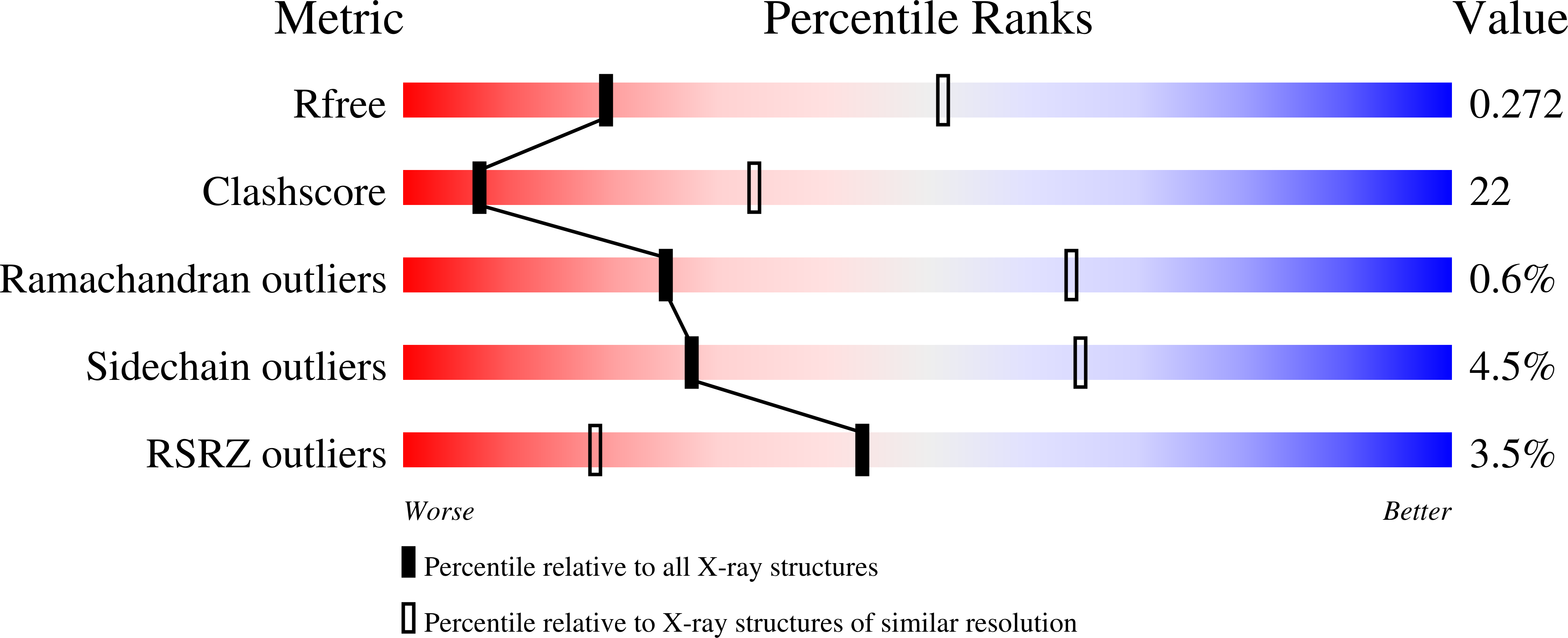
R-Value Free:
0.28
R-Value Work:
0.24
Space Group:
P 1 21 1
Macromolecular Entities
Polymer Type:polypeptide(L)
Molecule:Endonuclease I
Gene (Uniprot):3
Chain IDs:A, B, C, D, E, F, G, H, I, J
Chain Length:132
Number of Molecules:10
Biological Source:Enterobacteria phage T7
Primary Citation
The structure of a fibril-forming sequence, NNQQNY, in the context of a globular fold.
Protein Sci. 17 1617 1623 (2008)
PMID: 18552127 DOI: 10.1110/ps.036368.108

Abstact

Numerous human disorders are associated with the formation of protein fibrils. The fibril-forming capacity of a protein has been found in recent studies to be determined by a short segment of residues that forms a dual beta-sheet, called a steric zipper, in the spine of the fibril. The question arises as to whether a fibril-forming segment, when inserted within the sequence of a globular protein, will invariably cause the protein to form fibrils. Here we investigate this question by inserting the known fibril-forming segment NNQQNY into the globular enzyme, T7 endonuclease I. From earlier studies, we know that in its fibril form, NNQQNY is in an extended conformation. We first found that the inserted NNQQNY stimulates fibril formation of T7 endonuclease I in solution. Thus NNQQNY within T7 endonuclease I can exist in an extended conformation, capable of forming the steric zipper in the core of a fibril. We also found that T7 endonuclease I folds into a decamer that does not form fibrils. We determined the structure of the decamer by X-ray crystallography, finding an unusual oligomer without point group symmetry, and finding that the NNQQNY segments within the decamer adopt two twisted conformations, neither is apparently able to fibrillize. We conclude that twisting of fibril forming sequences from the fully extended conformation, imposed by the context of their placement in proteins, can interfere with fibril formation.

Legend

Protein

Chemical

Disease

Primary Citation of related structures
Feedback Form
Name
Email
Institute
Feedback