3AXX image
Deposition Date 2011-04-18
Release Date 2012-02-01
Last Version Date 2024-10-23
Entry Detail
PDB ID:
3AXX
Keywords:
Title:
Functional analysis of hyperthermophilic endocellulase from the Archaeon Pyrococcus horikoshii
Biological Source:
Source Organism(s):
Expression System(s):
Method Details:
Experimental Method:
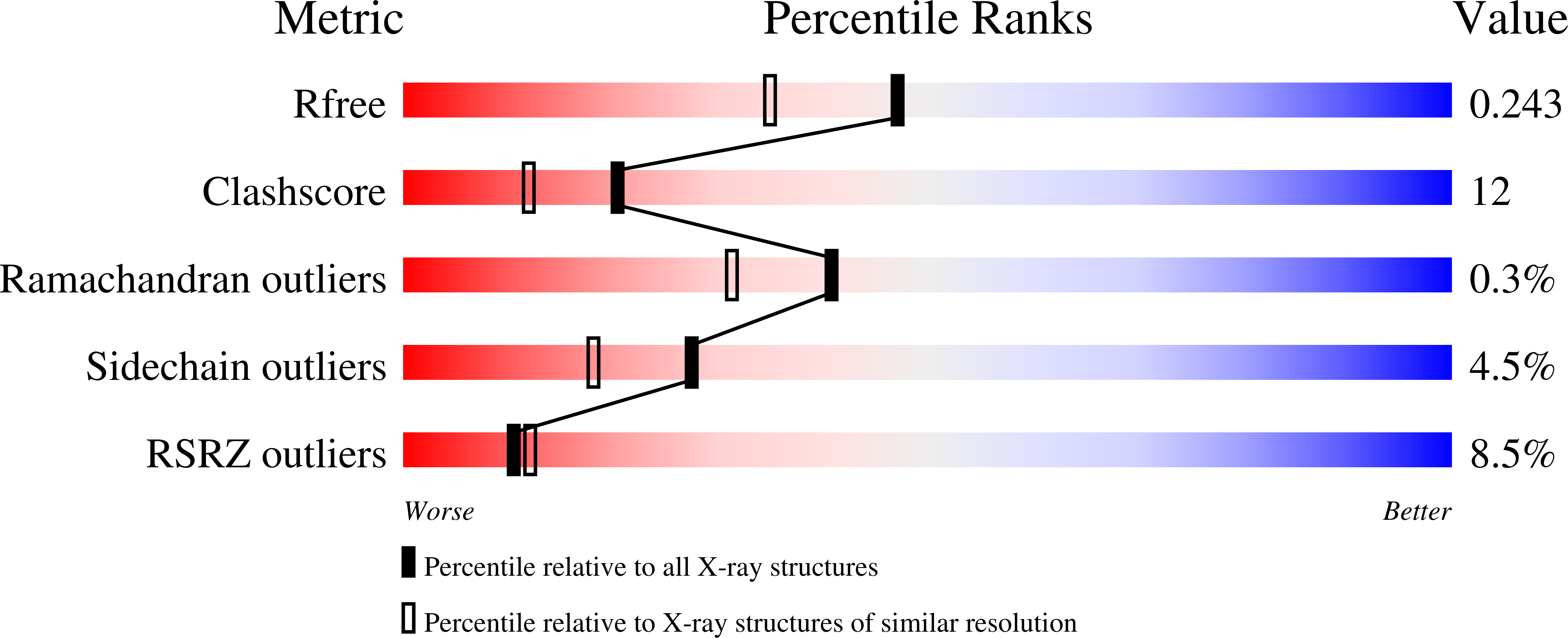
Resolution:
1.90 Å
R-Value Free:
0.24
R-Value Work:
0.22
Space Group:
C 1 2 1
Macromolecular Entities
Structures with similar UniProt ID
Protein Blast
Polymer Type:polypeptide(L)
Molecule:458aa long hypothetical endo-
Gene (Uniprot):PH1171
Chain IDs:A, B, C
Chain Length:458
Number of Molecules:3
Biological Source:Pyrococcus horikoshii
Ligand Molecules
Peptide-like Molecules
PRD_900005
Primary Citation
Functional analysis of hyperthermophilic endocellulase from Pyrococcus horikoshii by crystallographic snapshots
Biochem. J. 437 223 230 (2011)
PMID: 21557724 DOI: 10.1042/BJ20110292

Abstact

A hyperthermophilic membrane-related β-1,4-endoglucanase (family 5, cellulase) of the archaeon Pyrococcus horikoshii was found to be capable of hydrolysing cellulose at high temperatures. The hyperthermophilic cellulase has promise for applications in biomass utilization. To clarify its detailed function, we determined the crystal structures of mutants of the enzyme in complex with either the substrate or product ligands. We were able to resolve different kinds of complex structures at 1.65-2.01 Å (1 Å=0.1 nm). The structural analysis of various mutant enzymes yielded a sequence of crystallographic snapshots, which could be used to explain the catalytic process of the enzyme. The substrate position is fixed by the alignment of one cellobiose unit between the two aromatic amino acid residues at subsites +1 and +2. During the enzyme reaction, the glucose structure of cellulose substrates is distorted at subsite -1, and the β-1,4-glucoside bond between glucose moieties is twisted between subsites -1 and +1. Subsite -2 specifically recognizes the glucose residue, but recognition by subsites +1 and +2 is loose during the enzyme reaction. This type of recognition is important for creation of the distorted boat form of the substrate at subsite -1. A rare enzyme-substrate complex was observed within the low-activity mutant Y299F, which suggested the existence of a trapped ligand structure before the formation by covalent bonding of the proposed intermediate structure. Analysis of the enzyme-substrate structure suggested that an incoming water molecule, essential for hydrolysis during the retention process, might be introduced to the cleavage position after the cellobiose product at subsites +1 and +2 was released from the active site.

Legend

Protein

Chemical

Disease

Primary Citation of related structures
Feedback Form
Name
Email
Institute
Feedback