

37VY image
Deposition Date 2026-08-09
Release Date 2026-10-14
Last Version Date 2026-10-14
Entry Detail
PDB ID:
37VY
Title:
CTE Type I-like structure
Biological Source:
Source Organism(s):
Homo sapiens (Taxon ID: 9606)
Method Details:
Experimental Method:
Resolution:
2.96 Å
Aggregation State:
FILAMENT
Reconstruction Method:
HELICAL
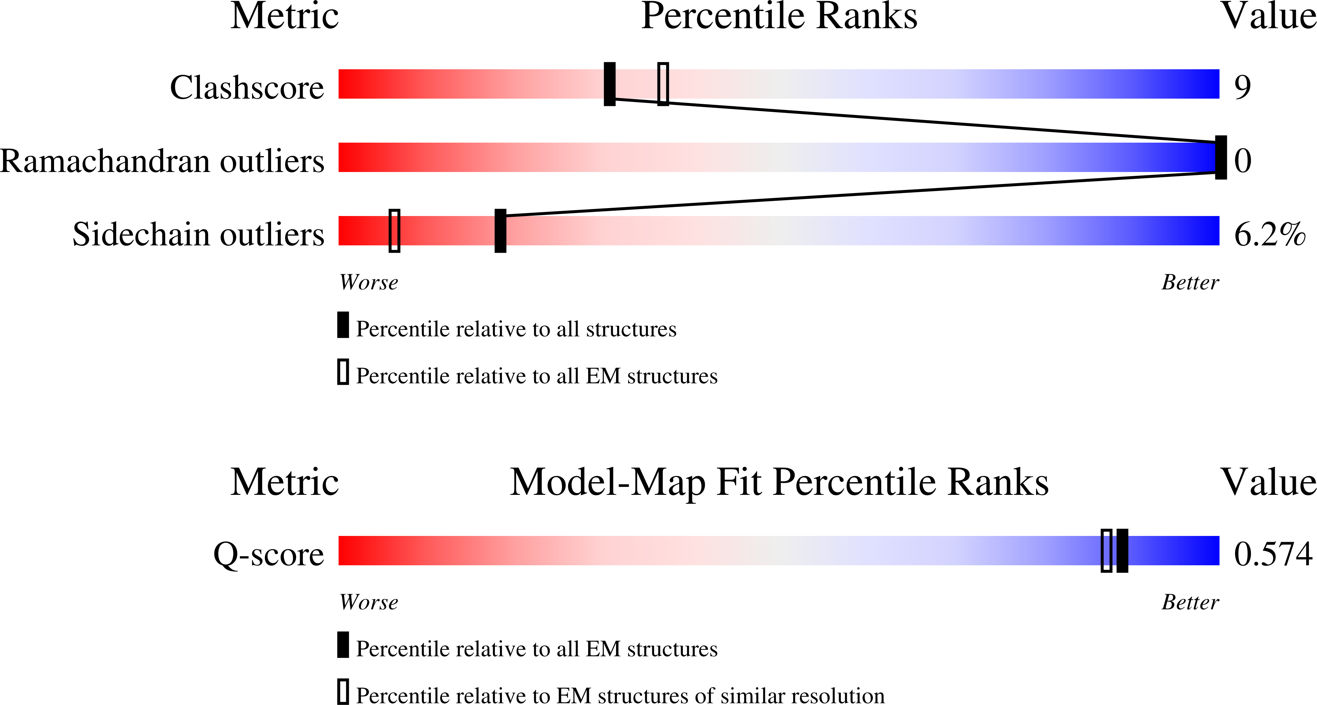
Macromolecular Entities
Polymer Type:polypeptide(L)
Molecule:Isoform Tau-F of Microtubule-
Chain IDs:A (auth: D), B (auth: F), C (auth: I), D (auth: B), E (auth: A), F (auth: G), G (auth: K), H (auth: L), I (auth: N), J (auth: E), K (auth: J), L (auth: C), M (auth: H), N (auth: M)
Chain Length:441
Number of Molecules:14
Biological Source:Homo sapiens
Ligand Molecules
Primary Citation
Structural heterogeneity of CTE Type I tau filaments with an interface-shifted polymorph.
Biochem.Biophys.Res.Commun. 838 154679 154679 (2026)
PMID: 42826653 DOI: 10.1016/j.bbrc.2026.154679

Abstact

Tau filament types can differ in inter-protofilament packing while sharing the same protofilament fold. Motivated by our recent findings in vacuolar tauopathy, we analyzed cryo-electron microscopy (cryo-EM) data from brain-derived chronic traumatic encephalopathy (CTE) tau filaments deposited in EMPIAR-10313. We resolved the canonical CTE Type I structure and a previously unreported CTE Type I-like structure with a shifted protofilament interface at 2.78 and 2.96 A, respectively. Mapping particle-state assignments back onto the original micrographs showed that the CTE Type I and CTE Type I-like packing states can occupy locally contiguous regions within the same fibrils. These findings identify an interface-shifted CTE Type I-like structure and support its local coexistence with CTE Type I within individual fibrils.

Legend

Protein

Chemical

Disease

Primary Citation of related structures
Feedback Form
Name
Email
Institute
Feedback