

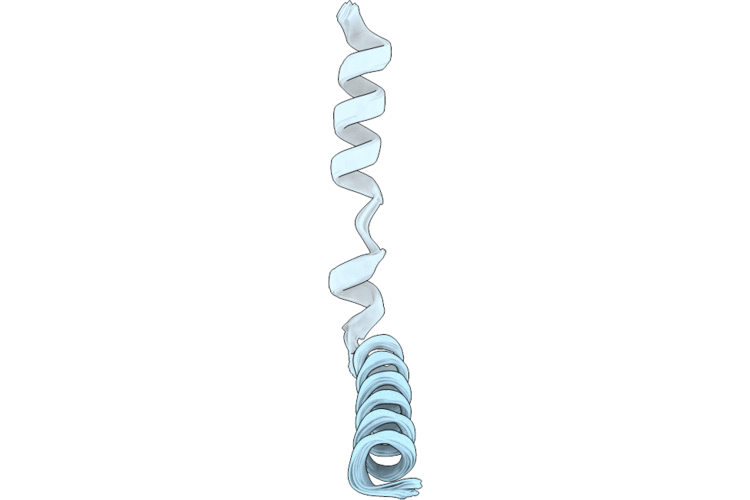
37CT image
Deposition Date 2026-07-14
Release Date 2026-08-26
Last Version Date 2026-08-26
Entry Detail
PDB ID:
37CT
Title:
Solid-State NMR Structure of FtsL, a Cell Division Protein from Mycobacterium tuberculosis, in Lipid Bilayers
Biological Source:
Source Organism(s):
Expression System(s):
Method Details:
Experimental Method:
Conformers Calculated:
14
Conformers Submitted:
10
Selection Criteria:
structures with the lowest energy
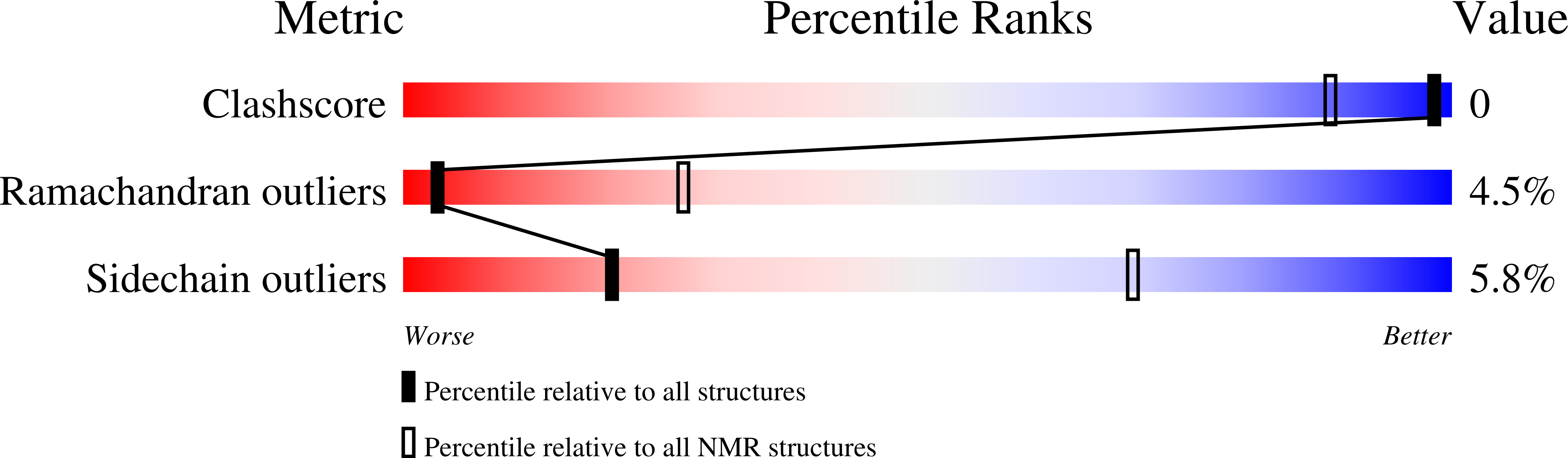
Macromolecular Entities
Polymer Type:polypeptide(L)
Molecule:Probable conserved proline ri
Gene (Uniprot):Rv2164c
Chain IDs:A
Chain Length:49
Number of Molecules:1
Biological Source:Mycobacterium tuberculosis H37Rv
Ligand Molecules
Primary Citation
Structure and Membrane Interactions of Two Helices from Mycobacterium tuberculosis FtsL, One Transmembrane and One Amphipathic.
J.Am.Chem.Soc. 148 33990 34001 (2026)
PMID: 42584571 DOI: 10.1021/jacs.6c12502

Abstact

Many transmembrane (TM) proteins contain an amphipathic helix. These amphipathic helices can sense lipid composition, stabilize membrane curvature, and mediate protein-protein interactions. Here, we combined solid-state NMR spectroscopy (ssNMR) and molecular dynamics simulations to characterize the structure and membrane interactions of a minimal TM and amphipathic construct (residues 101-146) from Mycobacterium tuberculosis (Mtb) FtsL. Amino-acid-specific 15N-labeled oriented-sample ssNMR spectra in POPC/POPG (4:1 molar ratio) membranes uniquely defined the orientation of the TM helix (residues 124-144), including a 16 degrees tilt, but constrained only the amphipathic helix (residues 101-114) to a 90 degrees tilt, leaving ambiguity in helical rotation and membrane burial depth. To resolve this ambiguity, we determined the two-dimensional free-energy surface using umbrella sampling simulations. The free-energy surface featured a major bound minimum, with the side chains of Leu104, Leu107, and Ile111 projecting into the hydrophobic core of the membrane and those of Arg103, Arg107, and Arg114 projecting sideways to interact with lipid headgroups. In pure POPC membranes, the bound basin contracted while the unbound basin expanded, as electrostatic attraction between Arg side chains and acidic POPG was replaced with repulsion by POPC's choline group. The final structure of FtsL101-146 was refined by restrained molecular dynamics simulations in a POPC/POPG bilayer and further validated by 13C-13C correlation magic-angle-spinning NMR. Together, these results demonstrate that the amphipathic helix of FtsL functions as a membrane-interacting element that stabilizes the protein in the membrane environment and mediates the recruitment of downstream proteins to the divisome for Mtb cell division.

Legend

Protein

Chemical

Disease

Primary Citation of related structures
Feedback Form
Name
Email
Institute
Feedback