

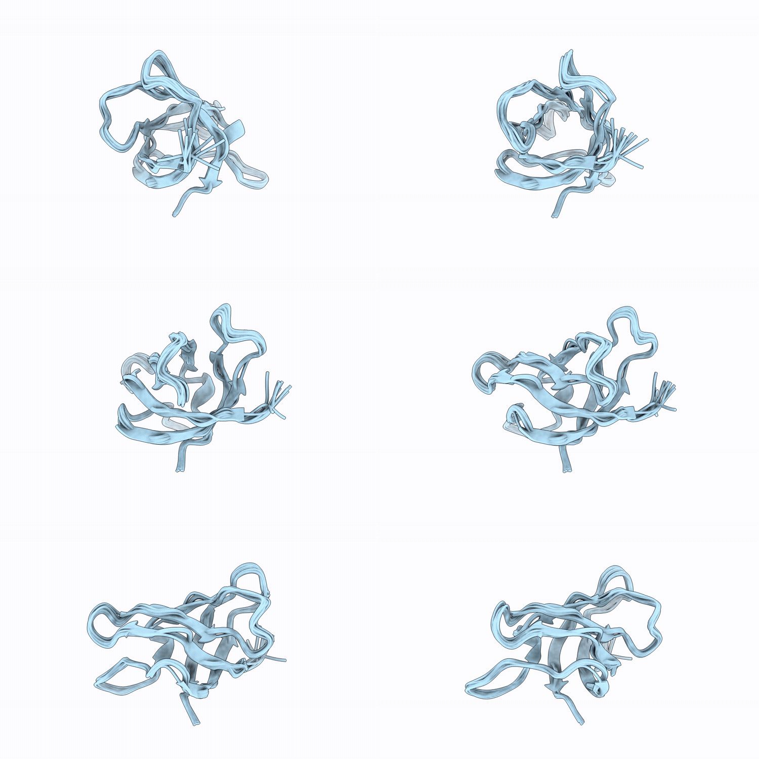
30IB image
Deposition Date 2026-04-28
Release Date 2026-05-27
Last Version Date 2026-05-27
Entry Detail
PDB ID:
30IB
Title:
NMR Solution Structure of Cold Shock Protein CspA
Biological Source:
Source Organism(s):
Escherichia coli (Taxon ID: 562)
Expression System(s):
Method Details:
Experimental Method:
Conformers Calculated:
100
Conformers Submitted:
10
Selection Criteria:
structures with the lowest energy

Macromolecular Entities
Structures with similar UniProt ID
Protein Blast
Polymer Type:polypeptide(L)
Molecule:Cold shock protein CspA
Gene (Uniprot):cspA
Chain IDs:A
Chain Length:70
Number of Molecules:1
Biological Source:Escherichia coli
Ligand Molecules
Primary Citation
Structural and functional insights into a mesophilic cold shock protein CspA with enhanced precision.
J.Magn.Reson. 388 108077 108077 (2026)
PMID: 42102648 DOI: 10.1016/j.jmr.2026.108077

Abstact

Cold-shock proteins (CSPs) are highly conserved nucleic acid-binding proteins that act as chaperones during cellular adaptation to low temperatures. Here, we present a comprehensive structural and dynamic characterization of Escherichia coli CspA using high-resolution NMR spectroscopy. The solution structure of CspA (PDB ID: 30IB) is supported by extensive NMR experimental restraints, minimal violations, and favorable stereochemistry, establishing it as a well-converged NMR structure. Crucially, we investigated backbone dynamics across multiple timescales, with a particular focus on the microsecond-millisecond regime using a combination of (15)N Carr-Purcell-Meiboom-Gill (CPMG) relaxation dispersion and (15)N chemical exchange saturation transfer (CEST) experiments, together with visible peak-position constraints. To our knowledge, this represents the first application of such a combined (15)N CEST, (15)N CPMG and visible peak-position constraints approach to probe conformational exchange in CSPs. Our results show that, in addition to the conserved aromatic residues of RNP1 and RNP2 motifs that mediate pi-stacking interactions with nucleic acids, an unexpectedly broad network of hydrophobic core and solvent-exposed polar residues undergoes conformational exchange. Notably, residues in the beta3-beta4 and beta4-beta5 loops display complex dynamics not fully captured by model-free analysis formalism, highlighting their role in binding site flexibility. Complementary AF3/YASARA modeling of the CspA bound to heptathymidine (dT7) further supported that aromatic and polar residues form pi-stacking and ionic interactions with ssDNA bases, corroborating the functional relevance of these dynamic regions. Therefore, our findings demonstrate that CspA relies on a dynamic network extending from conserved motifs through the hydrophobic core and flexible loops, conferring the structural adaptability required for efficient nucleic acid recognition and chaperone activity.

Legend

Protein

Chemical

Disease

Primary Citation of related structures
Feedback Form
Name
Email
Institute
Feedback