

2lw9 image
Deposition Date 2012-07-25
Release Date 2012-09-26
Last Version Date 2024-05-01
Entry Detail
PDB ID:
2LW9
Keywords:
Title:
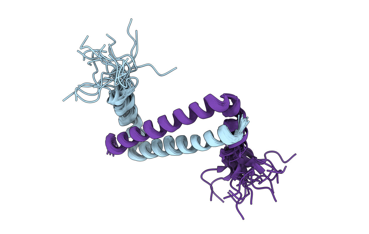
NMR solution structure of Myo10 anti-CC
Biological Source:
Source Organism(s):
Homo sapiens (Taxon ID: 9606)
Expression System(s):
Method Details:
Experimental Method:
Conformers Calculated:
61
Conformers Submitted:
20
Selection Criteria:
structures with the lowest energy
Macromolecular Entities
Polymer Type:polypeptide(L)
Molecule:Unconventionnal myosin-X
Gene (Uniprot):MYO10
Chain IDs:A, B
Chain Length:51
Number of Molecules:2
Biological Source:Homo sapiens
Ligand Molecules
Primary Citation
Antiparallel coiled-coil-mediated dimerization of myosin X
Proc. Natl. Acad. Sci. U.S.A. 109 17388 17393 (2012)
PMID: 23012428 DOI: 10.1073/pnas.1208642109

Abstact

Processive movements of unconventional myosins on actin filaments generally require motor dimerization. A commonly accepted myosin dimerization mechanism is via formation of a parallel coiled-coil dimer by a stretch of amino acid residues immediately carboxyl-terminal to the motor's lever-arm domain. Here, we discover that the predicted coiled-coil region of myosin X forms a highly stable, antiparallel coiled-coil dimer (anti-CC). Disruption of the anti-CC either by single-point mutations or by replacement of the anti-CC with a parallel coiled coil with a similar length compromised the filopodial induction activity of myosin X. We further show that the anti-CC and the single α-helical domain of myosin X are connected by a semirigid helical linker. The anti-CC-mediated dimerization may enable myosin X to walk on both single and bundled actin filaments.

Legend

Protein

Chemical

Disease

Primary Citation of related structures
Feedback Form
Name
Email
Institute
Feedback