2gm4 image
Deposition Date 2006-04-05
Release Date 2006-06-27
Last Version Date 2024-11-20
Entry Detail
PDB ID:
2GM4
Title:
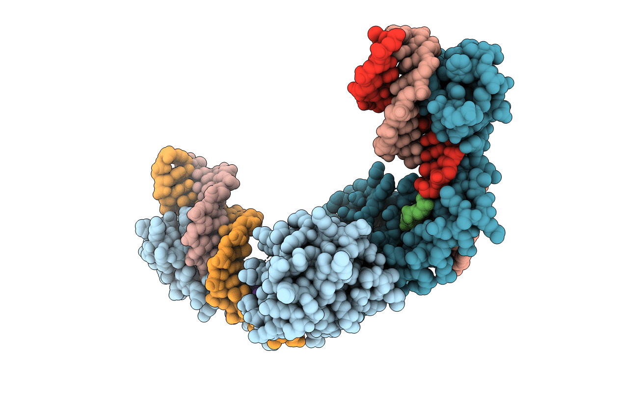
An activated, tetrameric gamma-delta resolvase: Hin chimaera bound to cleaved DNA
Biological Source:
Source Organism(s):
Escherichia coli (Taxon ID: 562)
Expression System(s):
Method Details:
Experimental Method:
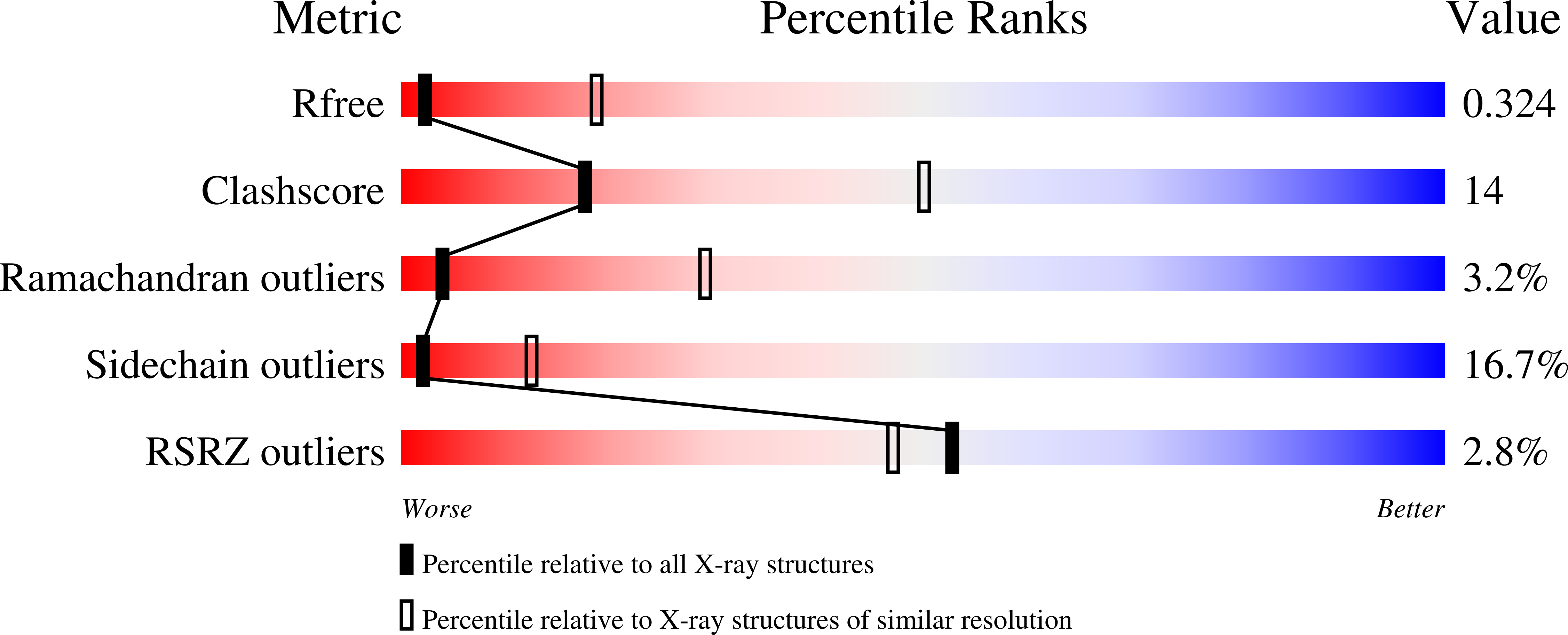
Resolution:
3.50 Å
R-Value Free:
0.32
R-Value Work:
0.28
R-Value Observed:
0.28
Space Group:
P 21 21 2
Macromolecular Entities
Structures with similar UniProt ID
Protein Blast
Polymer Type:polypeptide(L)
Molecule:Transposon gamma-delta resolv
Gene (Uniprot):tnpR
Mutagens:R2A, E56K, G96S, S98D, D100S, G101S, E102A, K105R, E124Q
Chain IDs:G (auth: A), H (auth: B)
Chain Length:183
Number of Molecules:2
Biological Source:Escherichia coli
Ligand Molecules
Primary Citation
Implications of structures of synaptic tetramers of gamma delta resolvase for the mechanism of recombination.
Proc. Natl. Acad. Sci. U.S.A. 103 10642 10647 (2006)
PMID: 16807292 DOI: 10.1073/pnas.0604062103

Abstact

The structures of two mutants of the site-specific recombinase, gammadelta resolvase, that form activated tetramers have been determined. One, at 3.5-A resolution, forms a synaptic intermediate of resolvase that is covalently linked to two cleaved DNAs, whereas the other is of an unliganded structure determined at 2.1-A resolution. Comparisons of the four known tetrameric resolvase structures show that the subunits interact through the formation of a common core of four helices. The N-terminal halves of these helices superimpose well on each other, whereas the orientations of their C termini are more variable. The catalytic domains of resolvase in the unliganded structure are arranged asymmetrically, demonstrating that their positions can move substantially while preserving the four-helix core that forms the tetramer. These results suggest that the precleavage synaptic tetramer of gammadelta resolvase, whose structure is not known, may be formed by a similar four-helix core, but differ in the relative orientations of its catalytic and DNA-binding domains.

Legend

Protein

Chemical

Disease

Primary Citation of related structures
Feedback Form
Name
Email
Institute
Feedback