

2dt2 image
Deposition Date 2006-07-09
Release Date 2006-07-25
Last Version Date 2024-11-06
Entry Detail
PDB ID:
2DT2
Title:
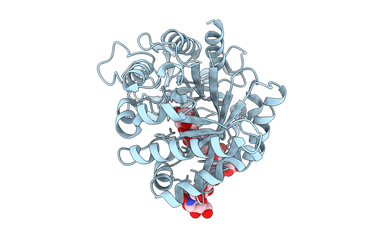
Crystal structure of the complex formed between goat signalling protein with pentasaccharide at 2.9A resolution
Biological Source:
Source Organism(s):
Capra hircus (Taxon ID: 9925)
Method Details:
Experimental Method:
Resolution:
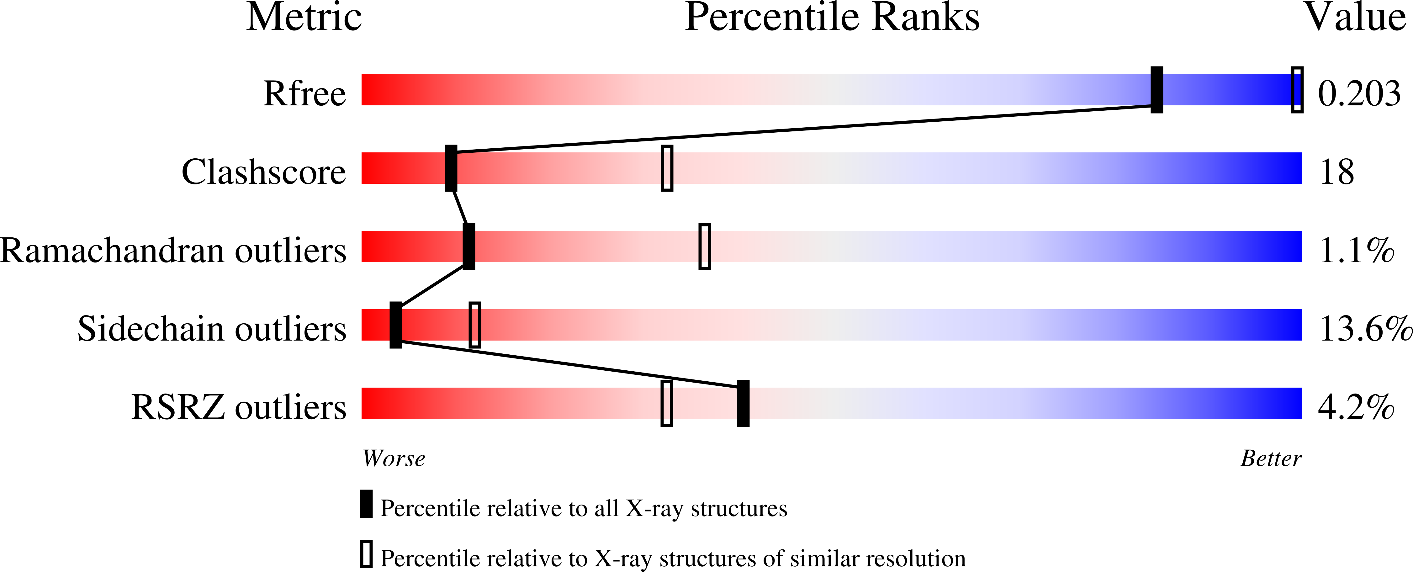
2.90 Å
R-Value Free:
0.20
R-Value Work:
0.19
R-Value Observed:
0.19
Space Group:
P 21 21 21
Macromolecular Entities
Polymer Type:polypeptide(L)
Molecule:Chitinase-3-like protein 1
Gene (Uniprot):CHI3L1
Chain IDs:A
Chain Length:361
Number of Molecules:1
Biological Source:Capra hircus
Modified Residue
Compound ID Chain ID Parent Comp ID Details 2D Image
ASN A ASN GLYCOSYLATION SITE
Primary Citation
Carbohydrate-binding properties of goat secretory glycoprotein (SPG-40) and its functional implications: structures of the native glycoprotein and its four complexes with chitin-like oligosaccharides
Acta Crystallogr. D Biol. Crystallogr. 63 437 446 (2007)
PMID: 17372347 DOI: 10.1107/S0907444907001631

Abstact

A 40 kDa glycoprotein (SPG-40) secreted during involution works as a protective signalling factor through its binding to viable cells. The crystal structure of the native protein has been determined at 2.3 A resolution. This is the first report on the carbohydrate-binding properties of SPG-40; the structure determinations of the complexes of SPG-40 with four oligosaccharides of different lengths at resolutions ranging from 2.2 to 2.8 A are described. Carbohydrate-binding studies with N-acetylglucosamines (GlcNAc(n), n = 3-6) using fluorescence spectroscopy revealed poor binding effects with GlcNAc(3) and GlcNAc(4), while GlcNAc(5) and GlcNAc(6) bound to SPG-40 with considerable strength; the dissociation constants (K(d)) were estimated to be 260 +/- 3 and 18 +/- 4 microM, respectively. SPG-40 was cocrystallized with GlcNAc(3), GlcNAc(4), GlcNAc(5) and GlcNAc(6). The overall structure of native SPG-40 was essentially similar to that reported previously at low resolution. The structures of its complexes with GlcNAc(3), GlcNAc(4), GlcNAc(5) and GlcNAc(6) revealed the positions of these oligosaccharides in the carbohydrate-binding groove and provided insights into the mechanism of binding of oligosaccharides to SPG-40, indicating that the preferred subsites in the carbohydrate-binding groove of SPG-40 were from -4 to -2. The structure of the protein remained unperturbed upon binding of GlcNAc(3) and GlcNAc(4), but the structure changed significantly upon binding of GlcNAc(5) and GlcNAc(6). Significant conformational variations were observed in the sugar-binding groove: Trp78 partially flipped out of the barrel in GlcNAc(5), while in the GlcNAc(6) complex a completely flipped-out Trp78 was observed along with several other conformational changes, including those of Asp186 and Arg242. Such changes upon binding to carbohydrates have not previously been observed in chitin-hydrolyzing chitinases and reflect less favourable binding of carbohydrates to SPG-40. As this appears to essentially be a binding protein, this loss of binding affinity might be compensated by other intermolecular interactions such as protein-protein interactions and also by the binding of its own glycan chain.

Legend

Protein

Chemical

Disease

Primary Citation of related structures
Feedback Form
Name
Email
Institute
Feedback