2Z2K image
Deposition Date 2007-05-22
Release Date 2007-07-24
Last Version Date 2023-11-01
Entry Detail
PDB ID:
2Z2K
Keywords:
Title:
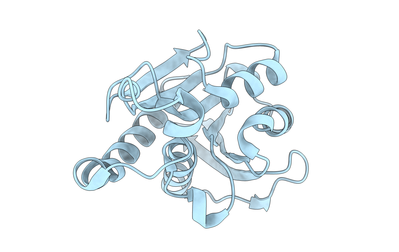
Crystal structure of Peptidyl-tRNA hydrolase from Mycobacterium tuberculosis
Biological Source:
Source Organism(s):
Expression System(s):
Method Details:
Experimental Method:
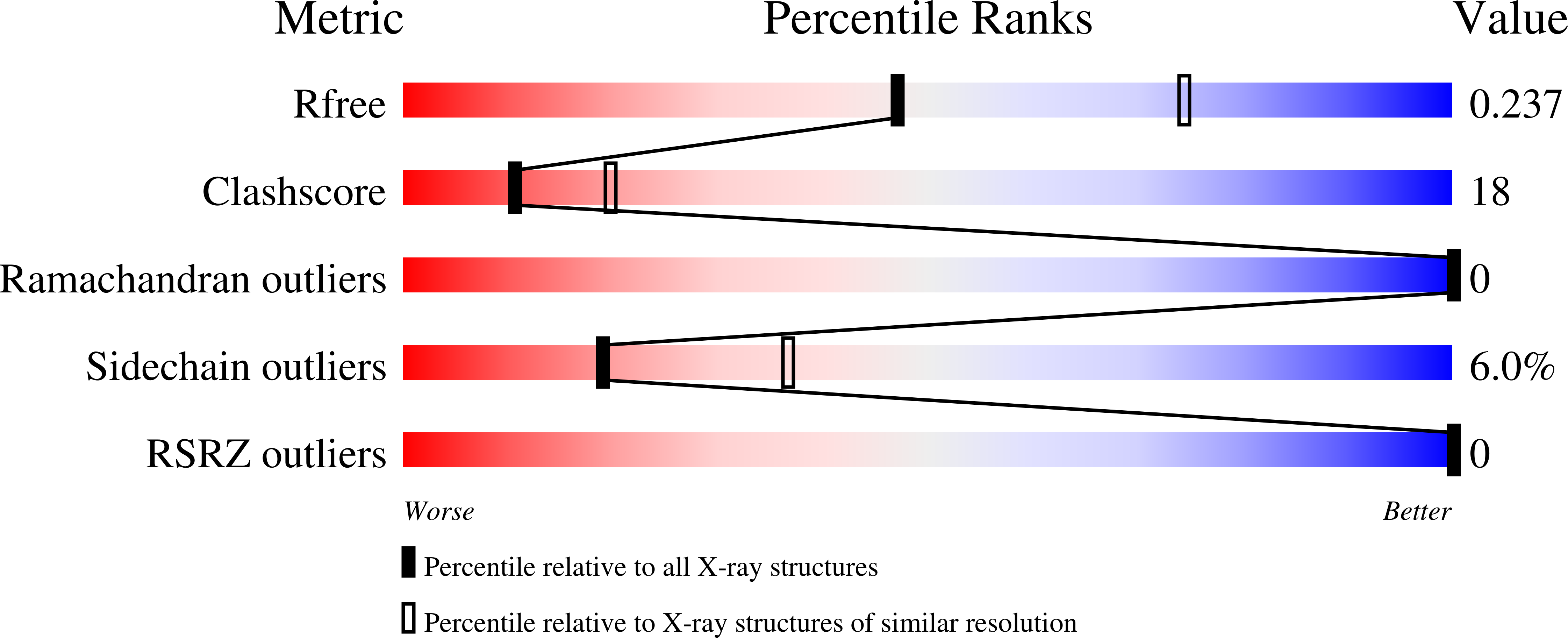
Resolution:
2.50 Å
R-Value Free:
0.24
R-Value Work:
0.19
R-Value Observed:
0.19
Space Group:
P 21 21 21
Macromolecular Entities
Structures with similar UniProt ID
Protein Blast
Polymer Type:polypeptide(L)
Molecule:Peptidyl-tRNA hydrolase
Chain IDs:A
Chain Length:191
Number of Molecules:1
Biological Source:Mycobacterium tuberculosis
Ligand Molecules
Primary Citation
Structural Plasticity and Enzyme Action: Crystal Structures of Mycobacterium tuberculosis Peptidyl-tRNA Hydrolase
J. Mol. Biol. 372 186 193 (2007)
PMID: 17619020 DOI: 10.1016/j.jmb.2007.06.053

Abstact

Peptidyl-tRNA hydrolase cleaves the ester bond between tRNA and the attached peptide in peptidyl-tRNA in order to avoid the toxicity resulting from its accumulation and to free the tRNA available for further rounds in protein synthesis. The structure of the enzyme from Mycobacterium tuberculosis has been determined in three crystal forms. This structure and the structure of the enzyme from Escherichia coli in its crystal differ substantially on account of the binding of the C terminus of the E. coli enzyme to the peptide-binding site of a neighboring molecule in the crystal. A detailed examination of this difference led to an elucidation of the plasticity of the binding site of the enzyme. The peptide-binding site of the enzyme is a cleft between the body of the molecule and a polypeptide stretch involving a loop and a helix. This stretch is in the open conformation when the enzyme is in the free state as in the crystals of M. tuberculosis peptidyl-tRNA hydrolase. Furthermore, there is no physical continuity between the tRNA and the peptide-binding sites. The molecule in the E. coli crystal mimics the peptide-bound enzyme molecule. The peptide stretch referred to earlier now closes on the bound peptide. Concurrently, a channel connecting the tRNA and the peptide-binding site opens primarily through the concerted movement of two residues. Thus, the crystal structure of M. tuberculosis peptidyl-tRNA hydrolase when compared with the crystal structure of the E. coli enzyme, leads to a model of structural changes associated with enzyme action on the basis of the plasticity of the molecule.

Legend

Protein

Chemical

Disease

Primary Citation of related structures
Feedback Form
Name
Email
Institute
Feedback