

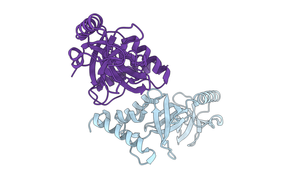
2WAC image
Deposition Date 2009-02-04
Release Date 2009-03-03
Last Version Date 2023-12-13
Entry Detail
PDB ID:
2WAC
Keywords:
Title:
Extended Tudor domain of Drosophila Melanogaster Tudor-SN (p100)
Biological Source:
Source Organism(s):
Expression System(s):
Method Details:
Experimental Method:
Resolution:
2.10 Å
R-Value Free:
0.26
R-Value Work:
0.22
R-Value Observed:
0.22
Space Group:
C 1 2 1

Macromolecular Entities
Polymer Type:polypeptide(L)
Molecule:CG7008-PA
Gene (Uniprot):Tudor-SN
Chain IDs:A, B
Chain Length:218
Number of Molecules:2
Biological Source:DROSOPHILA MELANOGASTER
Primary Citation
Structure and Ligand Binding of the Extended Tudor Domain of D. Melanogaster Tudor-Sn
J. Mol. Biol. 387 921 ? (2009)
PMID: 19232356 DOI: 10.1016/J.JMB.2009.02.018

Abstact

The Tudor-SN protein (p100, SND1) has been implicated in a variety of cellular processes, such as transcription, processing of edited double-stranded RNA, and splicing regulation. Molecular details of these functions are not yet understood. Tudor domains have previously been shown to bind methylated ligands, such as methylated lysines and arginines. It has been suggested that the role of Tudor-SN in splicing may involve binding to such methylated ligands or to the methylated 5' cap of spliceosomal snRNAs. Here, we report the crystal structure of the extended Tudor domain of Tudor-SN from Drosophila melanogaster to a resolution of 2.1 A. NMR secondary chemical shifts, relaxation data, and residual dipolar couplings indicate that the solution and crystal structures are similar. Binding of various ligands was investigated by NMR. Binding sites and affinities were characterized by chemical shift perturbations. We show that the aromatic cage of the Tudor domain specifically binds a peptide containing symmetrically dimethylated arginines (sDMA) with micromolar affinity, while the same peptide comprising nonmethylated arginines does not show significant chemical shift perturbations. Tudor-SN preferentially recognizes sDMA over asymmetrically dimethylated arginine (aDMA). In contrast, two 5' cap analogues with different methylation patterns, as well as mono-, di-, and trimethyllysines, show no binding. Our data demonstrate that the Tudor domain of Tudor-SN specifically recognizes sDMA-containing ligands. The aromatic cage of Tudor-SN is very similar to the one in the Tudor domain of the survival of motor neuron protein, which also recognizes sDMA peptides, indicating a conserved binding motif for this methylation mark. Recognition of sDMA in the C-terminal tails of spliceosomal Sm proteins suggests how Tudor-SN may interact with small nuclear ribonucleoprotein particles during the regulation of splicing.

Legend

Protein

Chemical

Disease

Primary Citation of related structures
Feedback Form
Name
Email
Institute
Feedback