2V25 image
Deposition Date 2007-06-01
Release Date 2007-06-26
Last Version Date 2023-12-13
Entry Detail
PDB ID:
2V25
Keywords:
Title:
Structure of the Campylobacter jejuni antigen Peb1A, an aspartate and glutamate receptor with bound aspartate
Biological Source:
Source Organism(s):
Expression System(s):
Method Details:
Experimental Method:
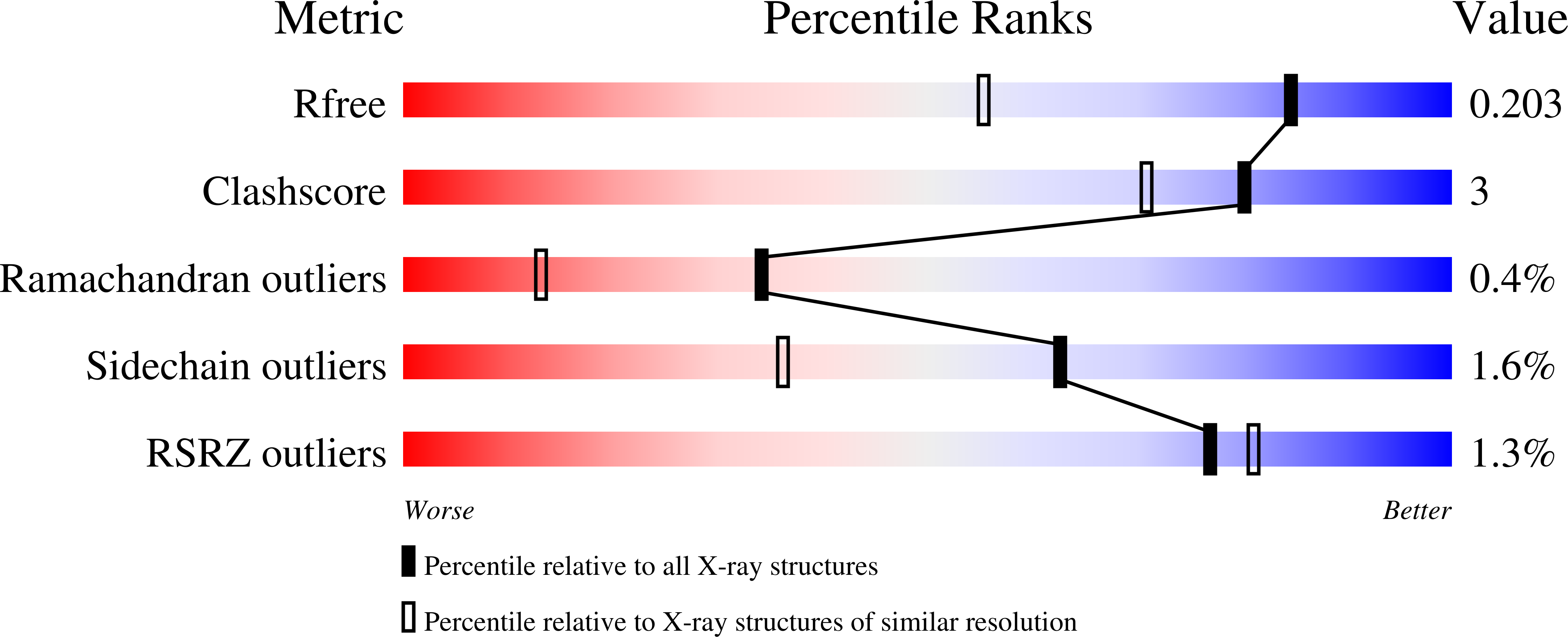
Resolution:
1.49 Å
R-Value Free:
0.18
R-Value Work:
0.16
R-Value Observed:
0.16
Space Group:
P 1 21 1
Macromolecular Entities
Structures with similar UniProt ID
Protein Blast
Polymer Type:polypeptide(L)
Molecule:MAJOR CELL-BINDING FACTOR
Gene (Uniprot):peb1A
Chain IDs:A, B
Chain Length:259
Number of Molecules:2
Biological Source:CAMPYLOBACTER JEJUNI
Primary Citation
A Bacterial Virulence Factor with a Dual Role as an Adhesin and a Solute Binding-Protein: The Crystal Structure at 1.5 A Resolution of the Peb1A Protein from the Food-Borne Human Pathogen Campylobacter Jejuni
J. Mol. Biol. 372 160 ? (2007)
PMID: 17631313 DOI: 10.1016/J.JMB.2007.06.041

Abstact

The PEB1a protein is an antigenic factor exposed on the surface of the food-borne human pathogen Campylobacter jejuni, which has a major role in adherence and host colonisation. PEB1a is also the periplasmic binding protein component of an aspartate/glutamate ABC transporter essential for optimal microaerobic growth on these dicarboxylic amino acids. Here, we report the crystal structure of PEB1a at 1.5 A resolution. The protein has a typical two-domain alpha/beta structure, characteristic of periplasmic extracytoplasmic solute receptors and a chain topology related to the type II subfamily. An aspartate ligand, clearly defined by electron density in the interdomain cleft, forms extensive polar interactions with the protein, the majority of which are made with the larger domain. Arg89 and Asp174 form ion-pairing interactions with the main chain alpha-carboxyl and alpha-amino-groups, respectively, of the ligand, while Arg67, Thr82, Lys19 and Tyr156 co-ordinate the ligand side-chain carboxyl group. Lys19 and Arg67 line a positively charged groove, which favours binding of Asp over the neutral Asn. The ligand-binding cleft is of sufficient depth to accommodate a glutamate. This is the first structure of an ABC-type aspartate-binding protein, and explains the high affinity of the protein for aspartate and glutamate, and its much weaker binding of asparagine and glutamine. Stopped-flow fluorescence spectroscopy indicates a simple bimolecular mechanism of ligand binding, with high association rate constants. Sequence alignments and phylogenetic analyses revealed PEB1a homologues in some Gram-positive bacteria. The alignments suggest a more distant homology with GltI from Escherichia coli, a known glutamate and aspartate-binding protein, but Lys19 and Tyr156 are not conserved in GltI. Our results provide a structural basis for understanding both the solute transport and adhesin/virulence functions of PEB1a.

Legend

Protein

Chemical

Disease

Primary Citation of related structures
Feedback Form
Name
Email
Institute
Feedback