

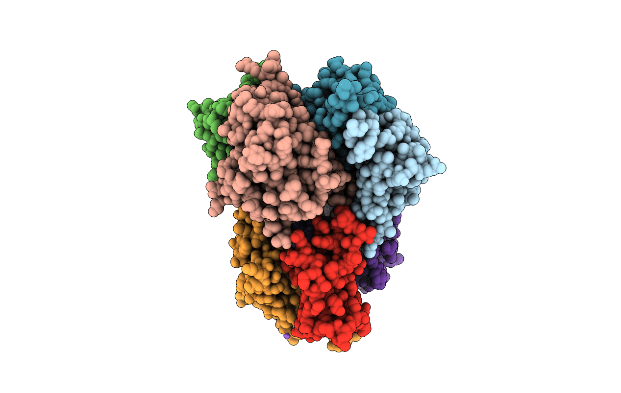
2RET image
Deposition Date 2007-09-27
Release Date 2008-02-05
Last Version Date 2024-10-30
Entry Detail
PDB ID:
2RET
Title:
The crystal structure of a binary complex of two pseudopilins: EpsI and EpsJ from the Type 2 Secretion System of Vibrio vulnificus
Biological Source:
Source Organism(s):
Expression System(s):
Method Details:
Experimental Method:
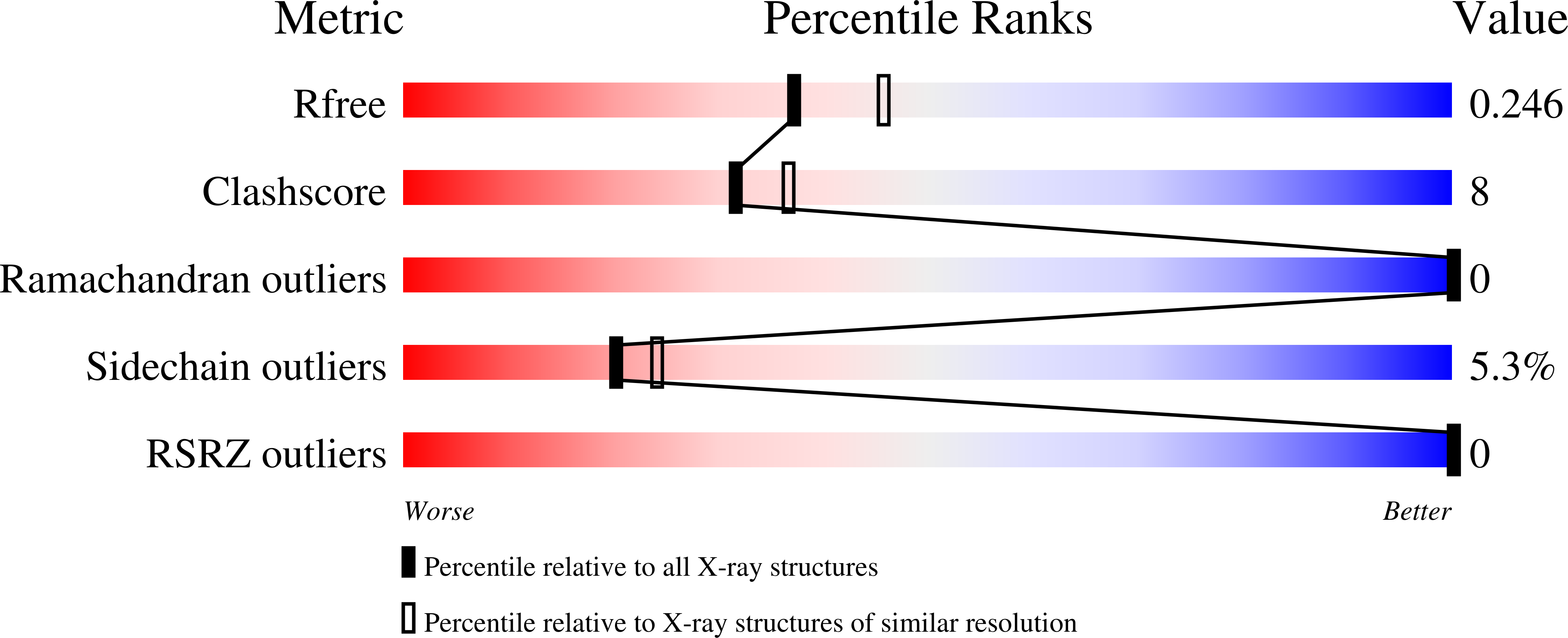
Resolution:
2.21 Å
R-Value Free:
0.24
R-Value Work:
0.18
R-Value Observed:
0.18
Space Group:
P 1
Macromolecular Entities
Polymer Type:polypeptide(L)
Molecule:Pseudopilin EpsI
Gene (Uniprot):VV0219
Mutagens:E128T, K129T
Chain IDs:A, C, E, G
Chain Length:103
Number of Molecules:4
Biological Source:Vibrio vulnificus
Polymer Type:polypeptide(L)
Molecule:EpsJ
Chain IDs:B, D, F, H
Chain Length:175
Number of Molecules:4
Biological Source:Vibrio vulnificus
Modified Residue
Compound ID Chain ID Parent Comp ID Details 2D Image
MSE A MET SELENOMETHIONINE
Primary Citation
The crystal structure of a binary complex of two pseudopilins: EpsI and EpsJ from the type 2 secretion system of Vibrio vulnificus.
J. Mol. Biol. 375 471 486 (2008)
PMID: 18022192 DOI: 10.1016/j.jmb.2007.10.035

Abstact

Type II secretion systems (T2SS) translocate virulence factors from the periplasmic space of many pathogenic bacteria into the extracellular environment. The T2SS of Vibrio cholerae and related species is called the extracellular protein secretion (Eps) system that consists of a core of multiple copies of 11 different proteins. The pseudopilins, EpsG, EpsH, EpsI, EpsJ and EpsK, are five T2SS proteins that are thought to assemble into a pseudopilus, which is assumed to interact with the outer membrane pore, and may actively participate in the export of proteins. We report here biochemical evidence that the minor pseudopilins EpsI and EpsJ from Vibrio species interact directly with one another. Moreover, the 2.3 A resolution crystal structure of a complex of EspI and EpsJ from Vibrio vulnificus represents the first atomic resolution structure of a complex of two different pseudopilin components from the T2SS. Both EpsI and EpsJ appear to be structural extremes within the family of type 4a pilin structures solved to date, with EpsI having the smallest, and EpsJ the largest, "variable pilin segment" seen thus far. A high degree of sequence conservation in the EpsI:EpsJ interface indicates that this heterodimer occurs in the T2SS of a large number of bacteria. The arrangement of EpsI and EpsJ in the heterodimer would correspond to a right-handed helical character of proteins assembled into a pseudopilus.

Legend

Protein

Chemical

Disease

Primary Citation of related structures
Feedback Form
Name
Email
Institute
Feedback