

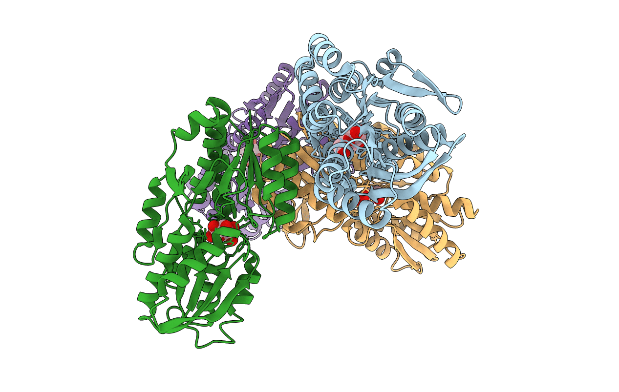
2QVC image
Deposition Date 2007-08-08
Release Date 2007-08-28
Last Version Date 2024-10-30
Entry Detail
PDB ID:
2QVC
Title:
Crystal structure of a periplasmic sugar ABC transporter from Thermotoga maritima
Biological Source:
Source Organism(s):
Expression System(s):
Method Details:
Experimental Method:
Resolution:
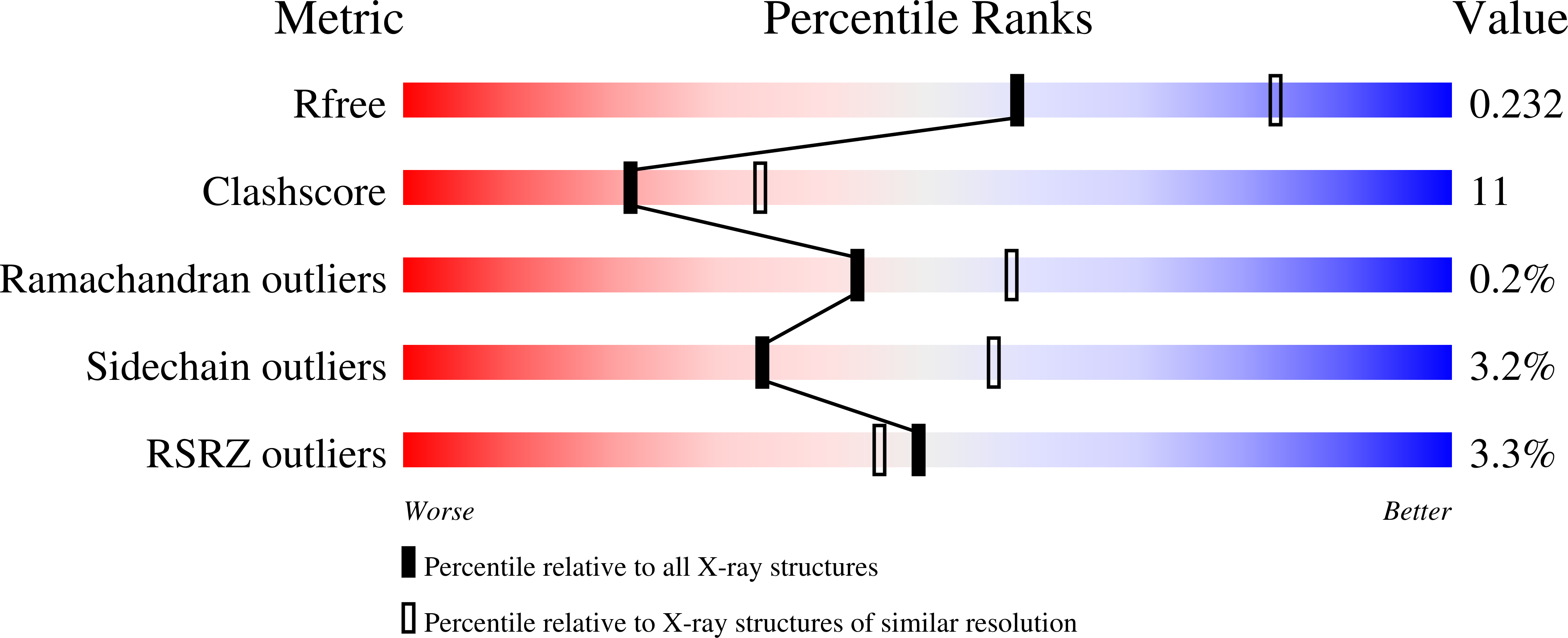
2.40 Å
R-Value Free:
0.24
R-Value Work:
0.21
R-Value Observed:
0.21
Space Group:
H 3
Macromolecular Entities
Polymer Type:polypeptide(L)
Molecule:Sugar ABC transporter, peripl
Gene (Uniprot):TM_0114
Chain IDs:A, B, C, D
Chain Length:313
Number of Molecules:4
Biological Source:Thermotoga maritima MSB8
Modified Residue
Compound ID Chain ID Parent Comp ID Details 2D Image
MSE A MET SELENOMETHIONINE
Ligand Molecules
Primary Citation
Structure of a periplasmic glucose-binding protein from Thermotoga maritima.
Acta Crystallogr. Sect. F Struct. Biol. Cryst. Commun. 68 1460 1464 (2012)
PMID: 23192024 DOI: 10.1107/S1744309112045241

Abstact

ABC transport systems have been characterized in organisms ranging from bacteria to humans. In most bacterial systems, the periplasmic component is the primary determinant of specificity of the transport complex as a whole. Here, the X-ray crystal structure of a periplasmic glucose-binding protein (GBP) from Thermotoga maritima determined at 2.4 Å resolution is reported. The molecule consists of two similar α/β domains connected by a three-stranded hinge region. In the current structure, a ligand (β-D-glucose) is buried between the two domains, which have adopted a closed conformation. Details of the substrate-binding sites revealed features that determine substrate specificity. In toto, ten residues from both domains form eight hydrogen bonds to the bound sugar and four aromatic residues (two from each domain) stabilize the substrate through stacking interactions.

Legend

Protein

Chemical

Disease

Primary Citation of related structures
Feedback Form
Name
Email
Institute
Feedback