2OE3 image
Deposition Date 2006-12-28
Release Date 2008-01-08
Last Version Date 2024-11-06
Entry Detail
PDB ID:
2OE3
Title:
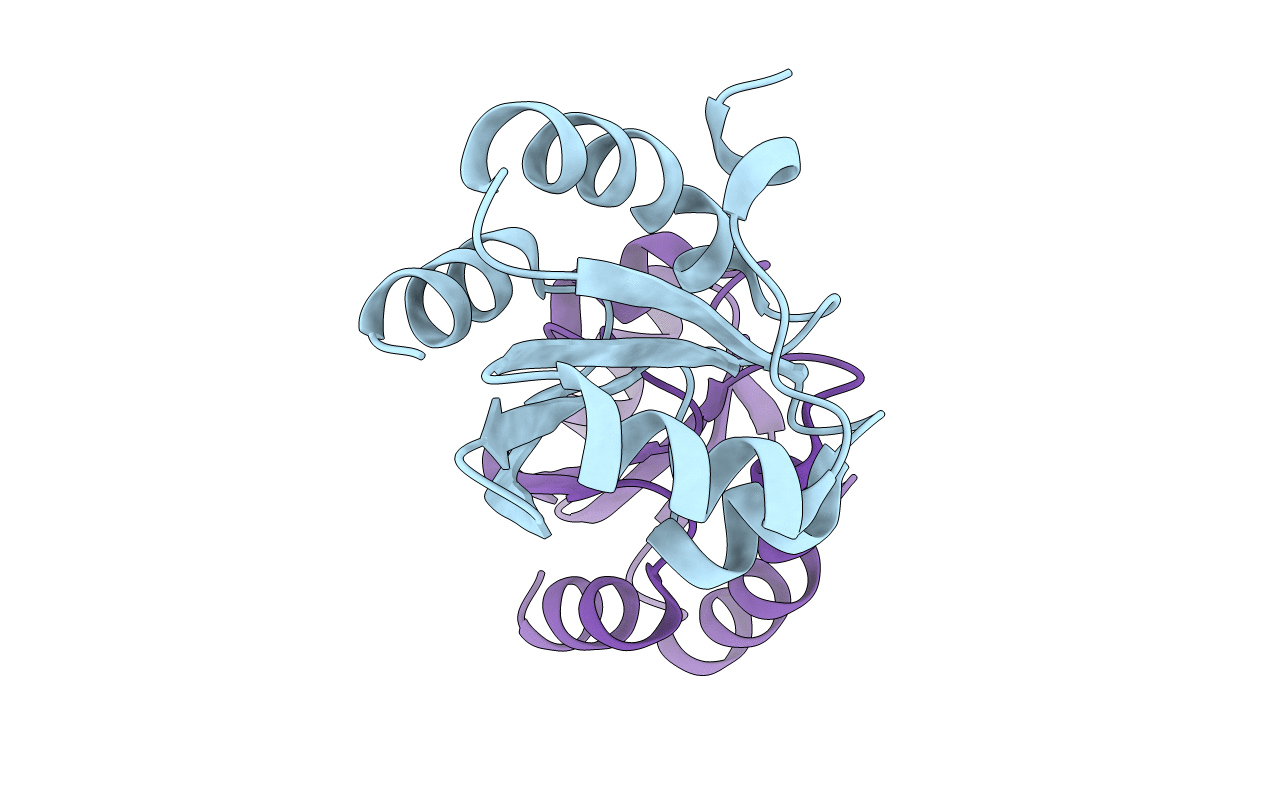
Crystal Structure of Mitochondrial Thioredoxin 3 from Saccharomyces cerevisiae (oxidized form)
Biological Source:
Source Organism(s):
Expression System(s):
Method Details:
Experimental Method:
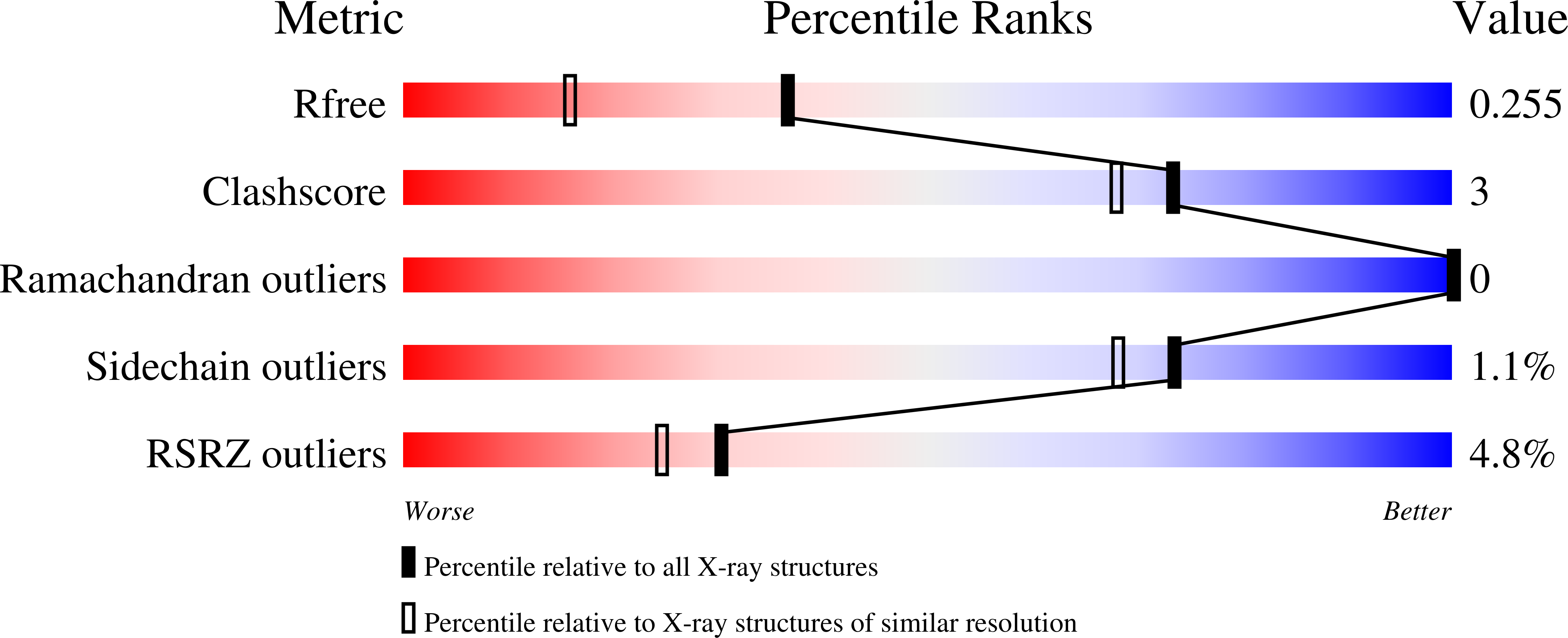
Resolution:
1.80 Å
R-Value Free:
0.25
R-Value Work:
0.19
R-Value Observed:
0.20
Space Group:
P 21 21 21
Macromolecular Entities
Structures with similar UniProt ID
Protein Blast
Polymer Type:polypeptide(L)
Molecule:Thioredoxin-3
Gene (Uniprot):TRX3
Chain IDs:A, B
Chain Length:114
Number of Molecules:2
Biological Source:Saccharomyces cerevisiae
Primary Citation
Structural and mechanistic analyses of yeast mitochondrial thioredoxin Trx3 reveal putative function of its additional cysteine residues
Biochim.Biophys.Acta 1794 716 721 (2009)
PMID: 19166985 DOI: 10.1016/j.bbapap.2008.12.016

Abstact

The yeast Saccharomyces cerevisiae Trx3 is a key member of the thioredoxin system to control the cellular redox homeostasis in mitochondria. We solved the crystal structures of yeast Trx3 in oxidized and reduced forms at 1.80 and 2.10 A, respectively. Besides the active site, the additional cysteine residue Cys69 also undergoes a significant redox-correlated conformational change. Comparative structural analyses in combination with activity assays revealed that residue Cys69 could be S-nitrosylated in vitro. S-nitrosylation of Cys69 will decrease the activity of Trx3 by 20%, which is comparable to the effect of the Cys69Ser mutation. Taken together, these findings provided us some new insights into the putative function of the additional cysteine residues of Trx3.

Legend

Protein

Chemical

Disease

Primary Citation of related structures
Feedback Form
Name
Email
Institute
Feedback