2LJH image
Deposition Date 2011-09-13
Release Date 2012-01-11
Last Version Date 2024-05-15
Entry Detail
PDB ID:
2LJH
Keywords:
Title:
NMR structure of Double-stranded RNA-specific editase Adar
Biological Source:
Source Organism(s):
Expression System(s):
Method Details:
Experimental Method:
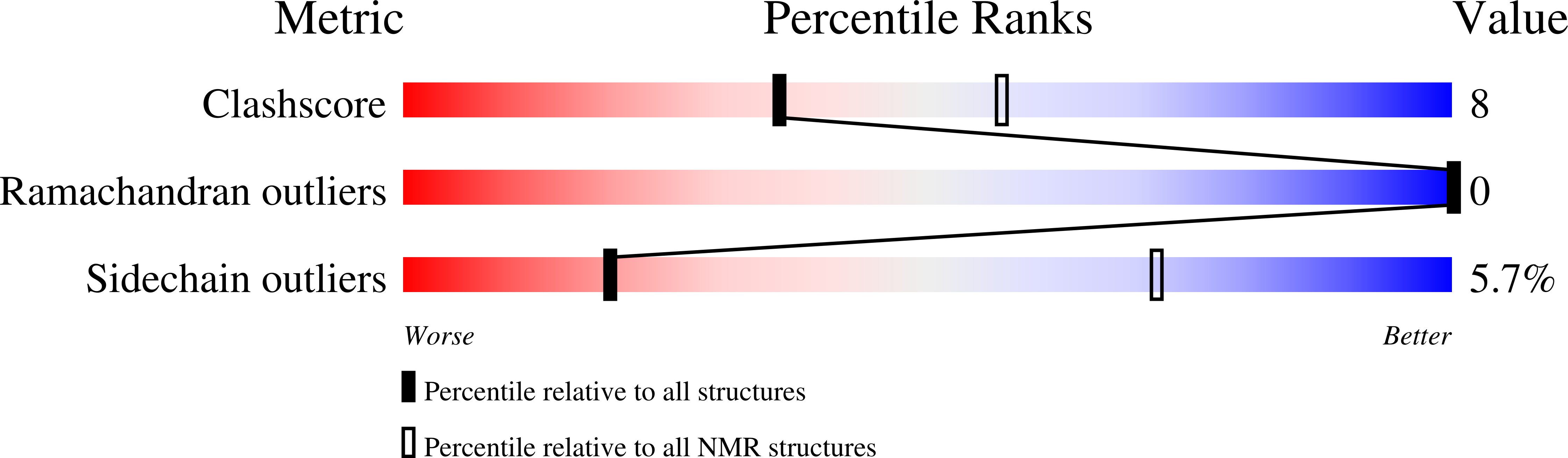
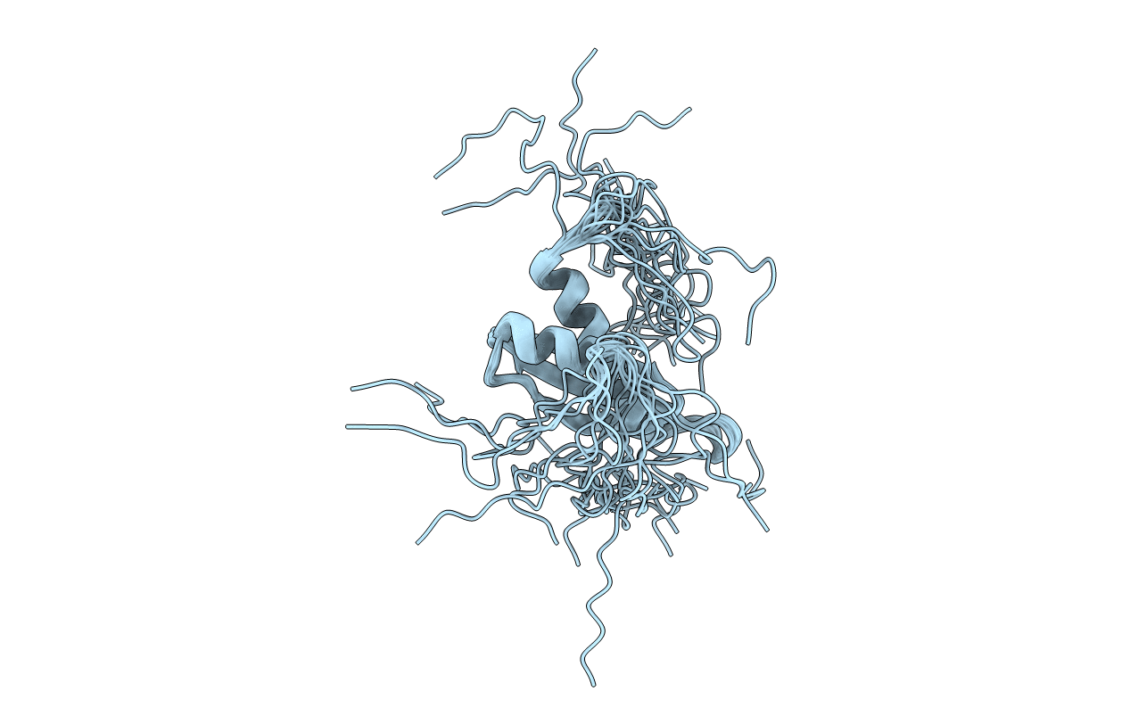
Conformers Calculated:
100
Conformers Submitted:
20
Selection Criteria:
structures with the lowest energy
Macromolecular Entities
Structures with similar UniProt ID
Protein Blast
Polymer Type:polypeptide(L)
Molecule:Double-stranded RNA-specific
Gene (Uniprot):Adar
Chain IDs:A
Chain Length:114
Number of Molecules:1
Biological Source:Drosophila melanogaster
Ligand Molecules
Primary Citation
Solution structure of the N-terminal dsRBD of Drosophila ADAR and interaction studies with RNA.
Biochimie 94 1499 1509 (2012)
PMID: 22210494 DOI: 10.1016/j.biochi.2011.12.017

Abstact

Adenosine deaminases that act on RNA (ADAR) catalyze adenosine to inosine (A-to-I) editing in double-stranded RNA (dsRNA) substrates. Inosine is read as guanosine by the translation machinery; therefore A-to-I editing events in coding sequences may result in recoding genetic information. Whereas vertebrates have two catalytically active enzymes, namely ADAR1 and ADAR2, Drosophila has a single ADAR protein (dADAR) related to ADAR2. The structural determinants controlling substrate recognition and editing of a specific adenosine within dsRNA substrates are only partially understood. Here, we report the solution structure of the N-terminal dsRNA binding domain (dsRBD) of dADAR and use NMR chemical shift perturbations to identify the protein surface involved in RNA binding. Additionally, we show that Drosophila ADAR edits the R/G site in the mammalian GluR-2 pre-mRNA which is naturally modified by both ADAR1 and ADAR2. We then constructed a model showing how dADAR dsRBD1 binds to the GluR-2 R/G stem-loop. This model revealed that most side chains interacting with the RNA sugar-phosphate backbone need only small displacement to adapt for dsRNA binding and are thus ready to bind to their dsRNA target. It also predicts that dADAR dsRBD1 would bind to dsRNA with less sequence specificity than dsRBDs of ADAR2. Altogether, this study gives new insights into dsRNA substrate recognition by Drosophila ADAR.

Legend

Protein

Chemical

Disease

Primary Citation of related structures
Feedback Form
Name
Email
Institute
Feedback