

2LCX image
Deposition Date 2011-05-11
Release Date 2012-05-16
Last Version Date 2024-05-15
Entry Detail
PDB ID:
2LCX
Keywords:
Title:
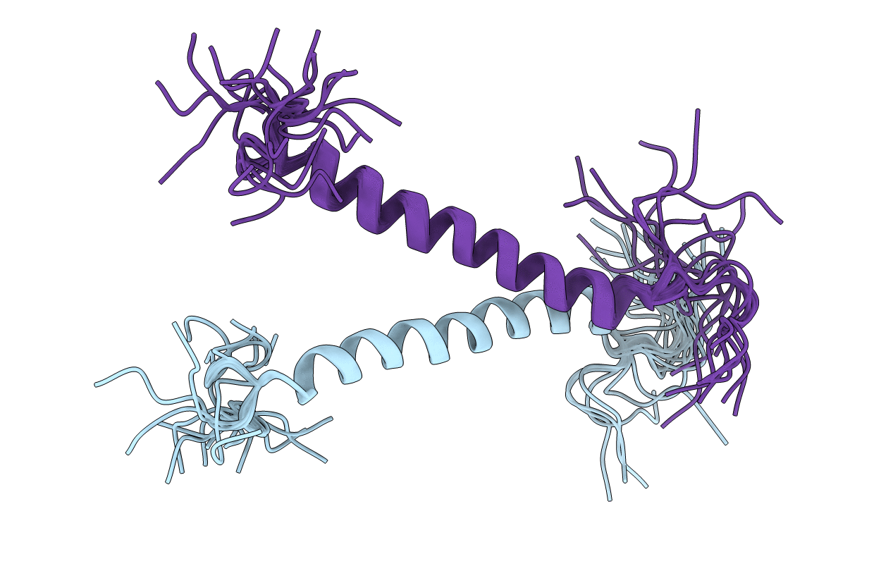
Spatial Structure of the ErbB4 dimeric TM domain
Biological Source:
Source Organism(s):
Homo sapiens (Taxon ID: 9606)
Expression System(s):
Method Details:
Experimental Method:
Conformers Calculated:
200
Conformers Submitted:
20
Selection Criteria:
target function
Macromolecular Entities
Structures with similar UniProt ID
Protein Blast
Polymer Type:polypeptide(L)
Molecule:Receptor tyrosine-protein kin
Gene (Uniprot):ERBB4
Chain IDs:A, B
Chain Length:44
Number of Molecules:2
Biological Source:Homo sapiens
Ligand Molecules
Primary Citation
Structural and thermodynamic insight into the process of "weak" dimerization of the ErbB4 transmembrane domain by solution NMR.
Biochim.Biophys.Acta 1818 2158 2170 (2012)
PMID: 22579757 DOI: 10.1016/j.bbamem.2012.05.001

Abstact

Specific helix-helix interactions between the single-span transmembrane domains of receptor tyrosine kinases are believed to be important for their lateral dimerization and signal transduction. Establishing structure-function relationships requires precise structural-dynamic information about this class of biologically significant bitopic membrane proteins. ErbB4 is a ubiquitously expressed member of the HER/ErbB family of growth factor receptor tyrosine kinases that is essential for the normal development of various adult and fetal human tissues and plays a role in the pathobiology of the organism. The dimerization of the ErbB4 transmembrane domain in membrane-mimicking lipid bicelles was investigated by solution NMR. In a bicellar DMPC/DHPC environment, the ErbB4 membrane-spanning α-helices (651-678)(2) form a right-handed parallel dimer through the N-terminal double GG4-like motif A(655)GxxGG(660) in a fashion that is believed to permit proper kinase domain activation. During helix association, the dimer subunits undergo a structural adjustment (slight bending) with the formation of a network of inter-monomeric polar contacts. The quantitative analysis of the observed monomer-dimer equilibrium provides insights into the kinetics and thermodynamics of the folding process of the helical transmembrane domain in the model environment that may be directly relevant to the process that occurs in biological membranes. The lipid bicelles occupied by a single ErbB4 transmembrane domain behave as a true ("ideal") solvent for the peptide, while multiply occupied bicelles are more similar to the ordered lipid microdomains of cellular membranes and appear to provide substantial entropic enhancement of the weak helix-helix interactions, which may be critical for membrane protein activity.

Legend

Protein

Chemical

Disease

Primary Citation of related structures
Feedback Form
Name
Email
Institute
Feedback