

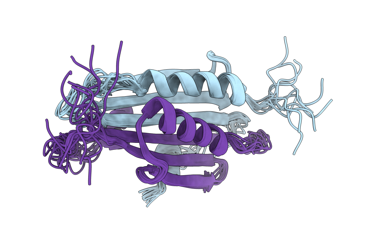
2KXO image
Deposition Date 2010-05-11
Release Date 2010-10-20
Last Version Date 2024-05-01
Entry Detail
PDB ID:
2KXO
Keywords:
Title:
Solution NMR structure of the cell division regulator MinE protein from Neisseria gonorrhoeae
Biological Source:
Source Organism(s):
Expression System(s):
Method Details:
Experimental Method:
Conformers Calculated:
200
Conformers Submitted:
10
Selection Criteria:
structures with the lowest energy
Macromolecular Entities
Structures with similar UniProt ID
Protein Blast
Polymer Type:polypeptide(L)
Molecule:Cell division topological spe
Gene (Uniprot):minE
Mutagens:E46A
Chain IDs:A, B
Chain Length:95
Number of Molecules:2
Biological Source:Neisseria gonorrhoeae
Ligand Molecules
Primary Citation
Appropriation of the MinD protein-interaction motif by the dimeric interface of the bacterial cell division regulator MinE.
Proc. Natl. Acad. Sci. U.S.A. 107 18416 18421 (2010)
PMID: 20937912 DOI: 10.1073/pnas.1007141107

Abstact

MinE is required for the dynamic oscillation of Min proteins that restricts formation of the cytokinetic septum to the midpoint of the cell in gram negative bacteria. Critical for this oscillation is MinD-binding by MinE to stimulate MinD ATP hydrolysis, a function that had been assigned to the first ∼30 residues in MinE. Previous models based on the structure of an autonomously folded dimeric C-terminal fragment suggested that the N-terminal domain is freely accessible for interactions with MinD. We report here the solution NMR structure of the full-length MinE dimer from Neisseria gonorrhoeae, with two parts of the N-terminal domain forming an integral part of the dimerization interface. Unexpectedly, solvent accessibility is highly restricted for residues that were previously hypothesized to directly interact with MinD. To delineate the true MinD-binding region, in vitro assays for MinE-stimulated MinD activity were performed. The relative MinD-binding affinities obtained for full-length and N-terminal peptides from MinE demonstrated that residues that are buried in the dimeric interface nonetheless participate in direct interactions with MinD. According to results from NMR spin relaxation experiments, access to these buried residues may be facilitated by the presence of conformational exchange. We suggest that this concealment of MinD-binding residues by the MinE dimeric interface provides a mechanism for prevention of nonspecific interactions, particularly with the lipid membrane, to allow the free diffusion of MinE that is critical for Min protein oscillation.

Legend

Protein

Chemical

Disease

Primary Citation of related structures
Feedback Form
Name
Email
Institute
Feedback