

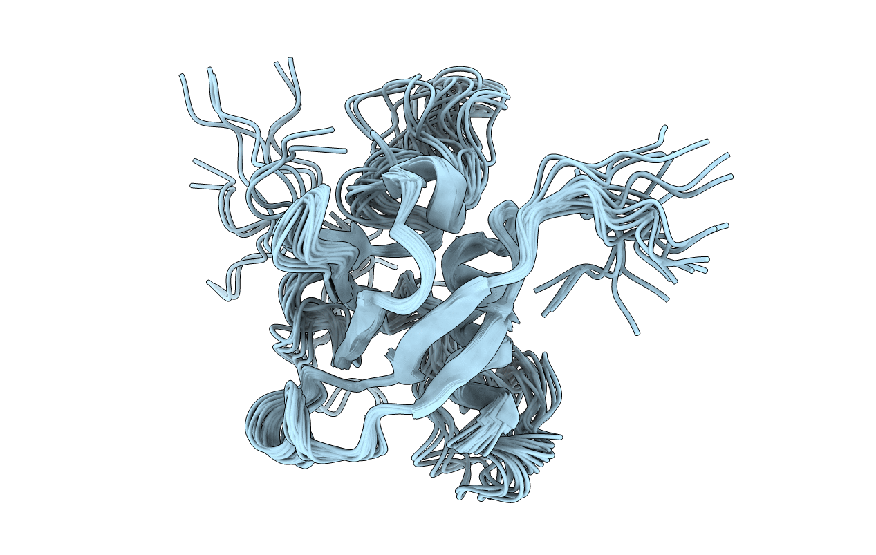
2JPH image
Deposition Date 2007-05-11
Release Date 2008-03-18
Last Version Date 2023-12-20
Entry Detail
PDB ID:
2JPH
Title:
NMR solution structure of the Rho GTPase binding domain of human plexin-b1
Biological Source:
Source Organism(s):
Homo sapiens (Taxon ID: 9606)
Expression System(s):
Method Details:
Experimental Method:
Conformers Calculated:
100
Conformers Submitted:
20
Selection Criteria:
structures with the least restraint violations

Macromolecular Entities
Polymer Type:polypeptide(L)
Molecule:Plexin-B1
Gene (Uniprot):PLXNB1
Mutagens:W1830F
Chain IDs:A
Chain Length:123
Number of Molecules:1
Biological Source:Homo sapiens
Ligand Molecules
Primary Citation
Insights into Oncogenic Mutations of Plexin-B1 Based on the Solution Structure of the Rho GTPase Binding Domain
Structure 16 246 258 (2008)
PMID: 18275816 DOI: 10.1016/j.str.2007.12.012

Abstact

The plexin family of transmembrane receptors are important for axon guidance, angiogenesis, but also in cancer. Recently, plexin-B1 somatic missense mutations were found in both primary tumors and metastases of breast and prostate cancers, with several mutations mapping to the Rho GTPase binding domain (RBD) in the cytoplasmic region of the receptor. Here we present the NMR solution structure of this domain, confirming that the protein has both a ubiquitin-like fold and surface features. Oncogenic mutations T1795A and T1802A are located in a loop region, perturb the average structure locally, and have no effect on Rho GTPase binding affinity. Mutations L1815F and L1815P are located at the Rho GTPase binding site and are associated with a complete loss of binding for Rac1 and Rnd1. Both are found to disturb the conformation of the beta3-beta4 sheet and the orientation of surrounding side chains. Our study suggests that the oncogenic behavior of the mutants can be rationalized with reference to the structure of the RBD of plexin-B1.

Legend

Protein

Chemical

Disease

Primary Citation of related structures
Feedback Form
Name
Email
Institute
Feedback