

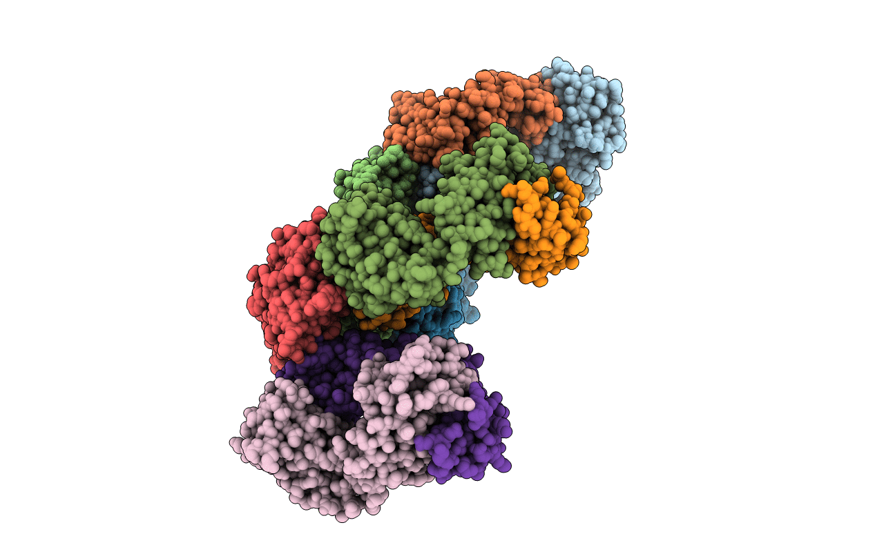
2JIX image
Deposition Date 2007-07-02
Release Date 2007-07-10
Last Version Date 2024-10-23
Entry Detail
PDB ID:
2JIX
Title:
Crystal structure of ABT-007 FAB fragment with the soluble domain of EPO receptor
Biological Source:
Source Organism(s):
HOMO SAPIENS (Taxon ID: 9606)
Expression System(s):
Method Details:
Experimental Method:
Resolution:
3.20 Å
R-Value Free:
0.32
R-Value Work:
0.25
R-Value Observed:
0.26
Space Group:
P 21 21 21

Macromolecular Entities
Polymer Type:polypeptide(L)
Molecule:ABT-007 FAB FRAGMENT
Chain IDs:A, G, I (auth: L)
Chain Length:214
Number of Molecules:3
Biological Source:HOMO SAPIENS
Polymer Type:polypeptide(L)
Molecule:ERYTHROPOIETIN RECEPTOR
Gene (Uniprot):EPOR
Chain IDs:B, C, E
Chain Length:225
Number of Molecules:3
Biological Source:HOMO SAPIENS
Polymer Type:polypeptide(L)
Molecule:ABT-007 FAB FRAGMENT
Chain IDs:D, F, H
Chain Length:217
Number of Molecules:3
Biological Source:HOMO SAPIENS
Ligand Molecules
Primary Citation
A Potent Erythropoietin-Mimicking Human Antibody Interacts Through a Novel Binding Site.
Blood 110 2408 ? (2007)
PMID: 17620453 DOI: 10.1182/BLOOD-2007-04-083998

Abstact

Recombinant human erythropoietin (rHu-EPO) is used to treat anemia by activating the erythropoietin receptor (EPOR) in erythroid progenitor cells, leading to proliferation and differentiation into mature red blood cells. To allow less frequent dosing, a hyperglycosylated version of EPO has been developed with a longer half-life. In principle, an agonistic antibody targeting EPOR would offer an even longer half-life, support robust monthly dosing, and, unlike EPO products, reduce the risk of pure red cell aplasia. The efficiency of signaling and corresponding potency of previously reported antibody mimics are generally suboptimal compared with EPO and not suitable for clinical use. Here we describe a potent, fully human, agonistic antibody (ABT007) targeting EPOR that supports potent, more sustained, and less pulsatile elevation of hematocrit in a human EPOR-expressing transgenic mouse model compared with standard doses of rHu-EPO while requiring less frequent dosing. Resolution of the crystal structure of the EPOR extracellular domain (ECD) complexed to the ABT007 Fab fragment, determined at 0.32 nm, identifies a binding site that is consistent with a novel mechanism of receptor activation based on a unique antibody-imposed conformational change. These results demonstrate that a symmetric molecule can serve as a potent activator of the EPOR.

Legend

Protein

Chemical

Disease

Primary Citation of related structures
Feedback Form
Name
Email
Institute
Feedback